. 2015 Apr;89(8):4356-71.
doi: 10.1128/JVI.03165-14. Epub 2015 Feb 4.

X-ray structure of the pestivirus NS3 helicase and its conformation in solution

Affiliations

X-ray structure of the pestivirus NS3 helicase and its conformation in solution

M Alejandra Tortorici et al. J Virol. 2015 Apr.

Abstract

Pestiviruses form a genus in the Flaviviridae family of small enveloped viruses with a positive-sense single-stranded RNA genome. Viral replication in this family requires the activity of a superfamily 2 RNA helicase contained in the C-terminal domain of nonstructural protein 3 (NS3). NS3 features two conserved RecA-like domains (D1 and D2) with ATPase activity, plus a third domain (D3) that is important for unwinding nucleic acid duplexes. We report here the X-ray structure of the pestivirus NS3 helicase domain (pNS3h) at a 2.5-Å resolution. The structure deviates significantly from that of NS3 of other genera in the Flaviviridae family in D3, as it contains two important insertions that result in a narrower nucleic acid binding groove. We also show that mutations in pNS3h that rescue viruses from which the core protein is deleted map to D3, suggesting that this domain may be involved in interactions that facilitate particle assembly. Finally, structural comparisons of the enzyme in different crystalline environments, together with the findings of small-angle X-ray-scattering studies in solution, show that D2 is mobile with respect to the rest of the enzyme, oscillating between closed and open conformations. Binding of a nonhydrolyzable ATP analog locks pNS3h in a conformation that is more compact than the closest apo-form in our crystals. Together, our results provide new insight and bring up new questions about pNS3h function during pestivirus replication.

Importance: Although pestivirus infections impose an important toll on the livestock industry worldwide, little information is available about the nonstructural proteins essential for viral replication, such as the NS3 helicase. We provide here a comparative structural and functional analysis of pNS3h with respect to its orthologs in other viruses of the same family, the flaviviruses and hepatitis C virus. Our studies reveal differences in the nucleic acid binding groove that could have implications for understanding the unwinding specificity of pNS3h, which is active only on RNA duplexes. We also show that pNS3h has a highly dynamic behavior--a characteristic probably shared with NS3 helicases from all Flaviviridae members--that could be targeted for drug design by using recent algorithms to specifically block molecular motion. Compounds that lock the enzyme in a single conformation or limit its dynamic range of conformations are indeed likely to block its helicase function.


Figures

FIG 1
FIG 1
Biochemical characterization of recombinant pNS3 and pNS3h from CSFV. (A) Schematic diagram of the pestivirus precursor polyprotein organization, indicating the individual mature proteins (boxes) and proteolytic processing sites (arrowheads or curved arrows for autoproteolytic cleavages). A diagram of the different constructs used in this study is shown below. (B) SDS-PAGE analysis of purified recombinant proteins stained with Coomassie blue. The lanes labeled 1 through 6 correspond to the various constructs listed in panel A. The middle lane displays prestained molecular mass markers. (C) The ATP hydrolysis activity of the proteins was measured by determination of the release of inorganic phosphate (y axis) and was plotted as a function of the ATP concentration (x axis). (D) Summary of the ATP hydrolysis properties of the different pNS3ph and pNS3h constructs. (E) The ATPase rate was measured in the presence of 0.2 mM ATP and the indicated concentrations of nonhydrolyzable ATP analogs (ADP·BeF3, AMPPcP, and ATPγS). Plotted is the amount of hydrolyzed ATP (y axis) against the ATP analog concentration (x axis). The standard deviations in panels C and E were estimated from two independent experiments. (F) The Km for ATP hydrolysis was determined by fitting the data from panel C with the Michaelis-Menten equation and is presented here in comparison to the corresponding values obtained for NS3 of other Flaviviridae members available in the literature. (G) The unwinding activities of pNS3ph and pNS3h were measured under multiple turnover assay conditions. The percentage of unwound RNA was calculated by dividing the amount of single-stranded RNA (RNA ss) by the total amount of RNA (single-stranded RNA plus double-stranded [RNA ds]). nd, not detected.
FIG 2
FIG 2
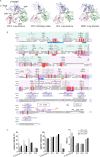
Structural comparison of NS3h orthologs. (A) Ribbon diagram of the pNS3h structure in its closed conformation from crystal form 2 (PDB accession no. 4CBG) (left), the homology model (http://www.bioacademy.gr/bioinformatics/csfv/Welcome.html) (second panel), HCV NS3h (PDB accession no. 1A1V) (third panel), and DENV-4 NS3h (PDB accession no. 2JLQ) (right). The proteins were superposed on D1 using the PDBefold program and are displayed in the same orientation. The three globular domains are colored green (D1), pink (D2), and blue (D3). (B) Structure-based alignment (produced using the Multalign and ESPript, version 2.2, programs) of pNS3h with its HCV and flavivirus counterparts. The CSFV (strain Alfort; GenBank accession no. J04358.2), HCV (strain H77; GenBank accession no. ACA48642), and DENV-4 (GenBank accession no. AAW30973) amino acid sequences of NS3h were retrieved from GenBank. The secondary structure elements of the CSFV, HCV, and DENV-4 NS3h proteins are displayed above the alignment. Identical or chemically similar residues are indicated at each position with a red background or red font, respectively. The residues in NS3 that allow the formation of core-less infectious pestivirus particles when substituted (described in Fig. 4) are highlighted in green. The residues deleted in the Δ1, Δ2, and Δ14 mutants (described in Fig. 3) are indicated by horizontal lines on top of the alignment. (C) The histograms show the percent amino acid sequence identity, the percentage of residues aligned, and the RMSD (Å) after the structure-based alignment between CSFV and HCV, CSFV and DENV, and HCV and DENV.
FIG 3
FIG 3
Domain 3 comparison with its NS3h orthologs. (A) Topology diagram of Flaviviridae NS3h domain 3. In pNS3h D3, the common core of three α helices shared with HCV and DENV NS3h is shown in blue, and the additional three α helices shared between CSFV and HCV D3 are shown in yellow. The secondary structure elements are numbered according to the structural alignment shown in Fig. 2. (B) Zoom view of CSFV NS3h D3. The α helices shared with HCV and DENV are shown using the same color code used for panel A. The two insertions in pNS3h D3, the L1 loop and helix α14, are colored orange. (C) Unwinding activity of pNS3, using construct pNS3p*h and the deletion mutants Δ1 and Δ2 (see Fig. 2B), measured using an RNA duplex template. The percentage of unwound RNA was calculated as described in the legend to Fig. 1.
FIG 4
FIG 4
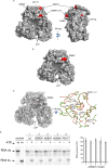
Mapping of the mutations in NS3 that allow formation of core-independent infectious pestivirus particles. (A) Surface representation of wild-type pNS3h1 recapitulating the locations of all the mutations (in red) that have individually been shown to rescue the viability of core-less CSFV particles. (B) Crystal structure of the pNS3h N588Y mutant in surface representation and in the same orientation as the wild-type enzyme in panel A. (C) (Left) Structure of the Q600K mutant. Because residue Q600 is buried, the surface is shown semitransparently. (Right) Zoom view of the Q600K mutant (the backbone is a green C-α trace; side chains are yellow sticks) superposed onto wild-type pNS3h (red, C-α trace; white sticks, side chains) as viewed from the ATP binding site (arrows in panels A and B). In wild-type pNS3h, the Q600 side chain makes a bidentate hydrogen bond with the E651 side chain (which is protonated at neutral pH, as it displays a pKa of 7.5). Both are represented as white sticks. In the Q600K mutant, the K600 side chain interacts in the same way with E651 (both side chains are shown as yellow sticks), but in this mutant it makes a salt bridge and compensates for the E651 negative charge (in this case, the pKa of E651 is 5.5). As the distances between the C-α atoms at positions 600 and 651 remain the same, the longer K600 side chain is in a slightly strained conformation. The side chain corresponding to other mutations with the same phenotype are also indicated in wild-type pNS3h (white sticks). (D) (Left) Unwinding activity of wild-type and mutant pNS3p*h. (Right) The bar graph represents the results of two independent RNA unwinding experiments made with two different preparations of proteins. The percentage of RNA unwound was calculated as described in the legend to Fig. 1.
FIG 5
FIG 5
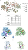
pNS3h and its conformational dynamics. (A) The crystallographic dimer observed in the various pNS3h crystal forms. pNS3h molecules within the AU of the crystals are related by a local 2-fold axis about which they make head-to-tail dimer interactions. This axis is perpendicular to the plane of the figure, and its intersection with this plane is represented here by a central black dot. The plot shows the 10 independent copies of the dimers superposed on the local 2-fold axes, showing that D1 and D3 superpose well but D2 has different orientations in each case. (B) Distances between the centers of mass (COM) of D1 and D2 (measured in Å). The distances were calculated using the same number of atoms for each domain in all molecules. The two columns of data correspond to the results for the two molecules within the crystallographic dimer displayed in panel A. In total, there are 16 protomers in which D2 was ordered and for which the distances between centers of mass could be determined. (C) The two conformations in crystal form 2. (Top) pNS3h in surface representation highlighting its characteristic arrowhead shape. Crystal form 2 displays the most open (left, light blue) and closed (right, pale green) conformations of pNS3h, shown here with D1 and D3 in the same orientation. D2 rotates by about 40 degrees as a rigid body around a hinge axis tilted by roughly 30 degrees with respect to the plane of the paper (the projection on the plane of the paper of the hinge axis is drawn as a thin black arrow). (Bottom) The same molecules from the top panels viewed down the hinge axis, with an X denoting the intersection of the hinge axis with the plane of the surface. The conserved helicase motifs are colored as described in the legend to Fig. 2B: cyan (H1 or Walker A), green (H1a), orange (H1b), red (H1c), pink (H2 or Walker B), light orange (H3), blue (H4), yellow (H4a), light purple (H5), and purple (H6). Motif Y, present only in the pesti- and hepacivirus genera, is shown in dark pink.
FIG 6
FIG 6
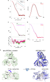
SAXS analysis. (A and B) Experimental scattering curves (A) and p(r) (B) of pNS3h in the apo-form (black curves) and in complex with ADP·BeF3 (red curves). (Inset in panel A) Guinier plots of the two samples. The regression lines are shown as cyan and gray, while the data used for the fit are indicated as black and red dots, respectively. The y axis of one data set was shifted vertically for clarity. a.u., arbitrary units. (C) The linear combination of the scattering curves derived from the conformations observed in crystal forms 2 and 3 together (magenta, χ2 = 2.0) fits the experimental scattering curve from apo-pNS3h (black) slightly better than the calculated scattering curve obtained with either of the intermediate forms of crystal form 3 (green, χ2 = 2.4), as indicated by the reduction of the corresponding residuals (bottom). (D) Experimental scattering curve of pNS3h in complex with ADP·BeF3 (red) superposed on the calculated scattering curve from the most closed conformation observed in crystal form 2 (purple), showing that the complex has a more compact conformation. (E) The remaining rearrangement necessary to generate the nucleotide-binding site in pNS3h. (Left) Apo-pNS3h in its closed conformation (pale green); (right) HCV NS3h in complex with ADP·BeF3 (PDB accession no. 3O8D) (blue). The structures were superposed on D1 and D3 using PDBfold. The D1-D2 linker is colored red in both structures. Dashed lines indicate disordered residues in the pNS3h structure. ADP·BeF3 and residues interacting with it in HCV NS3h are shown as orange and yellow sticks, respectively. The equivalent residues in pNS3h are also displayed in the same way, to show that a rotation of D2 of about 37 degrees (blue arrow) would be necessary to bring R506 and R509 into superposition with their HCV counterparts that bind the nucleotide, while displacing the D1-D2 linker out of the way.

Similar articles

Cited by

References

    1. Rossi S, Toigo C, Hars J, Pol F, Hamann JL, Depner K, Le Potier MF. 2011. New insights on the management of wildlife diseases using multi-state recapture models: the case of classical swine fever in wild boar. PLoS One 6:e24257. doi:10.1371/journal.pone.0024257. - DOI - PMC - PubMed
    1. Lindenbach BM, Murray CL,Thiel HJ, Rice CM. 2013. Flaviviridae, p 712–746. Knipe DM, Howley PM, Cohen JI, Griffin DE, Lamb RA, Martin MA, Racaniello VR, Roizman B (ed), Fields virology, 6th ed Lippincott Williams & Wilkins, Philadelphia, PA.
    1. Burbelo PD, Dubovi EJ, Simmonds P, Medina JL, Henriquez JA, Mishra N, Wagner J, Tokarz R, Cullen JM, Iadarola MJ, Rice CM, Lipkin WI, Kapoor A. 2012. Serology-enabled discovery of genetically diverse hepaciviruses in a new host. J Virol 86:6171–6178. doi:10.1128/JVI.00250-12. - DOI - PMC - PubMed
    1. Kapoor A, Simmonds P, Cullen JM, Scheel TK, Medina JL, Giannitti F, Nishiuchi E, Brock KV, Burbelo PD, Rice CM, Lipkin WI. 2013. Identification of a pegivirus (GB virus-like virus) that infects horses. J Virol 87:7185–7190. doi:10.1128/JVI.00324-13. - DOI - PMC - PubMed
    1. Lamp B, Riedel C, Roman-Sosa G, Heimann M, Jacobi S, Becher P, Thiel HJ, Rumenapf T. 2011. Biosynthesis of classical swine fever virus nonstructural proteins. J Virol 85:3607–3620. doi:10.1128/JVI.02206-10. - DOI - PMC - PubMed

Publication types

LinkOut - more resources