. 2015 Apr;89(8):4421-33.
doi: 10.1128/JVI.03619-14. Epub 2015 Feb 4.

Fine mapping and characterization of the L-polymerase-binding domain of the respiratory syncytial virus phosphoprotein

Affiliations

Fine mapping and characterization of the L-polymerase-binding domain of the respiratory syncytial virus phosphoprotein

Julien Sourimant et al. J Virol. 2015 Apr.

Abstract

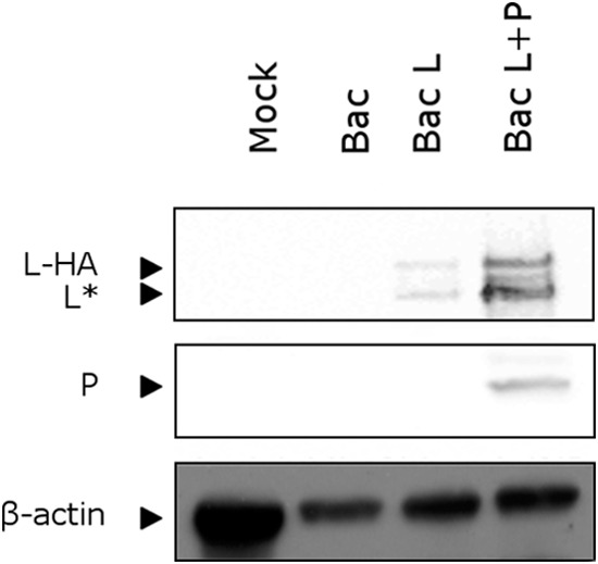
The minimum requirement for an active RNA-dependent RNA polymerase of respiratory syncytial virus (RSV) is a complex made of two viral proteins, the polymerase large protein (L) and the phosphoprotein (P). Here we have investigated the domain on P that is responsible for this critical P-L interaction. By use of recombinant proteins and serial deletions, an L binding site was mapped in the C-terminal region of P, just upstream of the N-RNA binding site. The role of this molecular recognition element of about 30 amino acid residues in the L-P interaction and RNA polymerase activity was evaluated in cellula using an RSV minigenome system and site-directed mutagenesis. The results highlighted the critical role of hydrophobic residues located in this region.

Importance: Respiratory syncytial virus (RSV) is the leading cause of lower respiratory tract illness in infants. Since no vaccine and no good antivirals against RSV are available, it is essential to better understand how the viral machinery functions in order to develop new antiviral strategies. Like all negative-strand RNA viruses, RSV codes for its own machinery to replicate and transcribe its genome. The core of this machinery is composed of two proteins, the phosphoprotein (P) and the large protein (L). Here, using recombinant proteins, we have mapped and characterized the P domain responsible for this L-P interaction and the formation of an active L-P complex. These findings extend our understanding of the mechanism of action of RSV RNA polymerase and allow us to define a new target for the development of drugs against RSV.


Figures

FIG 1
FIG 1
Expression of recombinant RSV HA-tagged L protein in insect cells. Recombinant baculoviruses expressing L-HA alone (Bac-L), coexpressing L-HA and P proteins (Bac-L/P), or expressing no foreign proteins (Bac) were used to infect Sf9 cells. At 72 h postinfection, cells were lysed, and the presence of L-HA and P in the soluble fraction was analyzed by Western blotting using anti-HA and anti-P antibodies. L* corresponds to the truncated form of L (residues 1 to 1789 of the original L sequence). A lysate from mock-infected Sf9 cells (Mock) was also included as a negative control. β-Actin was used as an internal standard. Western blots were imaged with a GeneGnome machine (Syngene).
FIG 2
FIG 2
A major L-binding domain is present in the C-terminal region of RSV P. (A) Schematic diagram of the GST-P fusion protein and of P deletion mutants used for P-L interaction studies. The names of the mutant GST-P proteins indicate the first and last remaining amino acids or deleted amino acids (GST-P[Δ122-160]), and the remaining sequences are shown as solid bars. The N-terminal (PNTD), oligomerization (POD), and C-terminal (PCTD) domains of P are indicated by arrows above the diagram. (B) P and truncated forms of P fused to GST and purified from bacteria on glutathione-Sepharose beads were incubated with lysates from Sf9 cells infected with the recombinant baculovirus expressing L-HA (Bac-L). After extensive washing, the presence or absence of L-HA was determined by SDS-PAGE and silver staining (top) or Western blotting (center) using anti-HA antibodies. The black star on the right of the silver-stained gel indicates contaminant proteins of higher molecular weight than L. GST-P proteins were visualized by SDS-PAGE and Coomassie blue staining (bottom).
FIG 3
FIG 3
Mapping the region of P involved in L binding by serial deletions. (A and B) Internal overlapping deletions of 20 to 30 (A) or 10 (B) amino acids were made throughout GST-PΔOD. (C) Stop codons were introduced between residues 232 and 241 of the original P sequence. The diagrams of the P deletion mutants used in these assays are shown to the left of the corresponding gels. Numbers indicate the first and the last residues of the deleted region of P. The presence of L-HA after GST pulldown was analyzed by Western blotting using anti-HA antibodies and exposure to X-ray film. GST-PΔOD-derived complexes were visualized by SDS-PAGE and Coomassie blue staining.
FIG 4
FIG 4
Effects of P point mutations on RSV polymerase activity. (A) Sequence alignments of the C-terminal region of P of the pneumoviruses human RSV (HRSV), bovine RSV (BRSV), ovine RSV (ORSV), pneumonia virus of mice (PVM), and canine pneumovirus (CPV) (GenBank accession numbers AAX23990.1, NP_048051.1, Q83956.1, Q5MKM7.1, and AHF88957.1, respectively) were made by Clustal Omega and prepared with ESPript 3. Two possible alignments between the most C-terminal residues of the different P proteins are shown. (B) Analysis of RSV polymerase activity with WT P and P substitution mutants. BSRT7/5 cells were transfected with RSV pP, pN, pL, and pM2-1 plasmids and an RSV-specific minigenome containing the firefly luciferase reporter gene, together with p-β-Gal, constitutively expressing β-galactosidase. Luciferase activity, measured 24 h after transfection, was normalized to β-galactosidase activity, and the luciferase activity gained with WT P was set to 100%. The mean values and confidence intervals (error bars) result from 3 separate experiments performed in triplicate. A negative control without P was run. (C) The expression of the different mutants was controlled in cell lysates by Western blotting using a polyclonal anti-P rabbit serum after 24 h of transfection and was compared to the expression of α-tubulin, used as an internal standard.
FIG 5
FIG 5
Point mutations affecting the P-L interaction in mammalian cells. (A) BSRT7/5 cells infected with MVA-T7 were transfected with pP (WT or mutant) and pL-HA plasmids. P proteins were immunoprecipitated (IP) from cell lysates 16 h after transfection by using a rabbit anti-P antiserum, and the presence of L-HA in the samples was analyzed by Western blotting using anti-HA antibodies. (Top) Cell lysates; (bottom) IP products. (B) Western blots were imaged with a GeneGnome machine (Syngene), and the L/P ratio was calculated before (filled bars) or after (shaded bars) immunoprecipitation. The L/P ratio is expressed as a percentage of the value obtained for WT P.
FIG 6
FIG 6
Effects of point mutations targeting the L-binding domain of P on the recruitment of L to cytoplasmic inclusion bodies. BSRT7/5 cells were transfected with pP, pN, and pL-EGFP plasmids. Cells were fixed 24 h after transfection, and the presence of L-EGFP in cytoplasmic inclusion bodies was observed by epifluorescence. The RSV N protein was revealed by immunofluorescence using rabbit polyclonal anti-N (1:500) and Alexa Fluor 594-conjugated goat anti-rabbit (1:2,000) antibodies. Nuclei were stained with Hoechst 33342 stain. Bars, 10 μm.
FIG 7
FIG 7
Helical-wheel projection of the L-binding domain of P, encompassing residues 216 to 234. (A) Summary of the effects of amino acid substitutions in the region of P encompassing amino acids 216 to 239 on RdRp activity, L-P interaction (as assessed by coimmunoprecipitation [CoIP]), and the presence of L in IBs. The amino acid sequence of the region of P encompassing amino acids 210 to 241 is given at the top. The nucleocapsid binding domain (NCBD) and L-binding domain (LBD) are indicated, and the predicted α-helix is represented. For RdRp activity, a minus sign, a plus-or-minus sign, and a plus sign indicate activities of ≤5%, ≤50%, and ≥50%, respectively. nd, not determined. (B) Assuming that the MoRE of P adopts a helical conformation upon the binding of L, we made a helical-wheel representation of this region using the HeliQuest program. This projection shows three leucines (L216, L223, and L227), which most likely constitute the direct interface for interaction with L, clustered on one side of the P molecular recognition helix. Hydrophobic residues are shown in yellow, negatively charged residues in red, and positively charged residues in blue. Red letters N and C indicate the N and C termini, respectively. Amino acids for which RdRp activity was reduced to <50% or <5% are labeled with one or two stars, respectively. The residue N224, replacement of which by Pro abrogated polymerase activity, is labeled with a number sign (#).

Similar articles

Cited by

References

    1. Tregoning JS, Schwarze J. 2010. Respiratory viral infections in infants: causes, clinical symptoms, virology, and immunology. Clin Microbiol Rev 23:74–98. doi:10.1128/CMR.00032-09. - DOI - PMC - PubMed
    1. Collins PL, Melero JA. 2011. Progress in understanding and controlling respiratory syncytial virus: still crazy after all these years. Virus Res 162:80–99. doi:10.1016/j.virusres.2011.09.020. - DOI - PMC - PubMed
    1. Valarcher JF, Taylor G. 2007. Bovine respiratory syncytial virus infection. Vet Res 38:153–180. doi:10.1051/vetres:2006053. - DOI - PubMed
    1. Loregian A, Marsden HS, Palu G. 2002. Protein-protein interactions as targets for antiviral chemotherapy. Rev Med Virol 12:239–262. doi:10.1002/rmv.356. - DOI - PubMed
    1. Morin B, Kranzusch PJ, Rahmeh AA, Whelan SP. 2013. The polymerase of negative-stranded RNA viruses. Curr Opin Virol 3:103–110. doi:10.1016/j.coviro.2013.03.008. - DOI - PMC - PubMed

MeSH terms

LinkOut - more resources