. 2015 Apr;89(8):4457-69.
doi: 10.1128/JVI.03612-14. Epub 2015 Feb 4.

Stress granule components G3BP1 and G3BP2 play a proviral role early in Chikungunya virus replication

Affiliations

Stress granule components G3BP1 and G3BP2 play a proviral role early in Chikungunya virus replication

Florine E M Scholte et al. J Virol. 2015 Apr.

Abstract

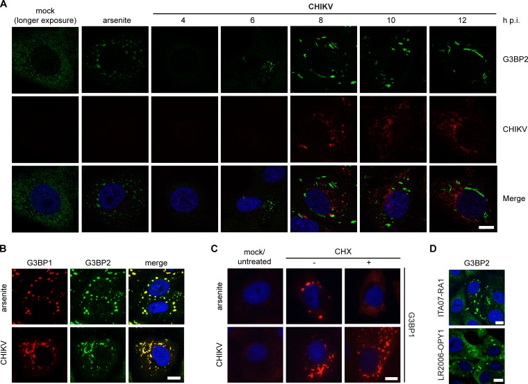
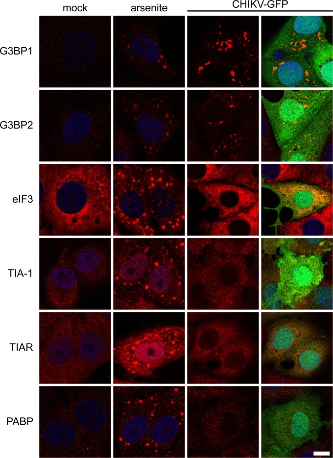
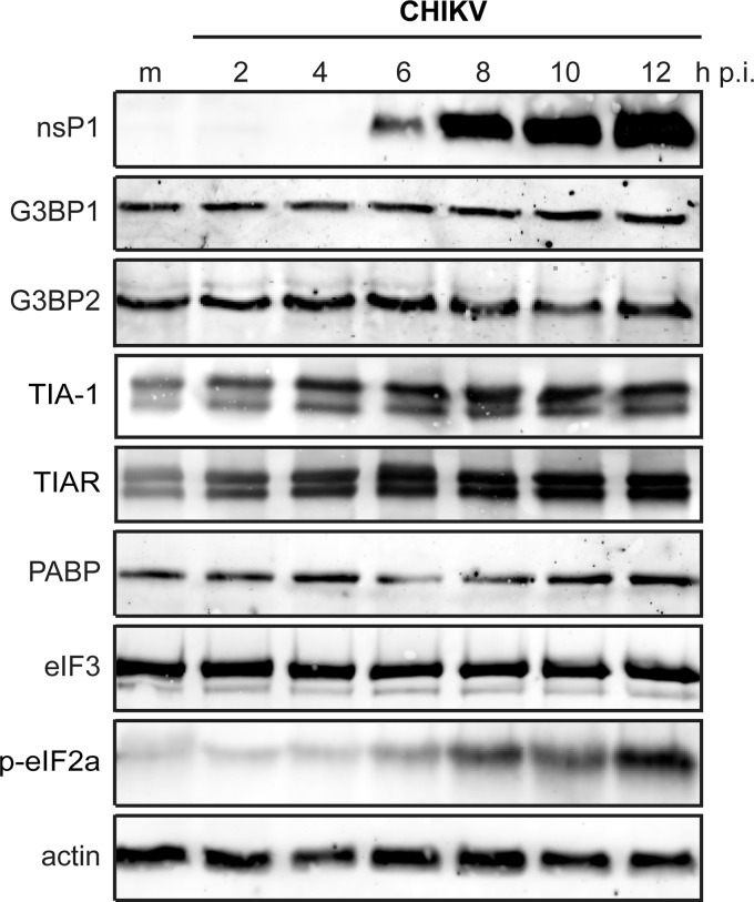
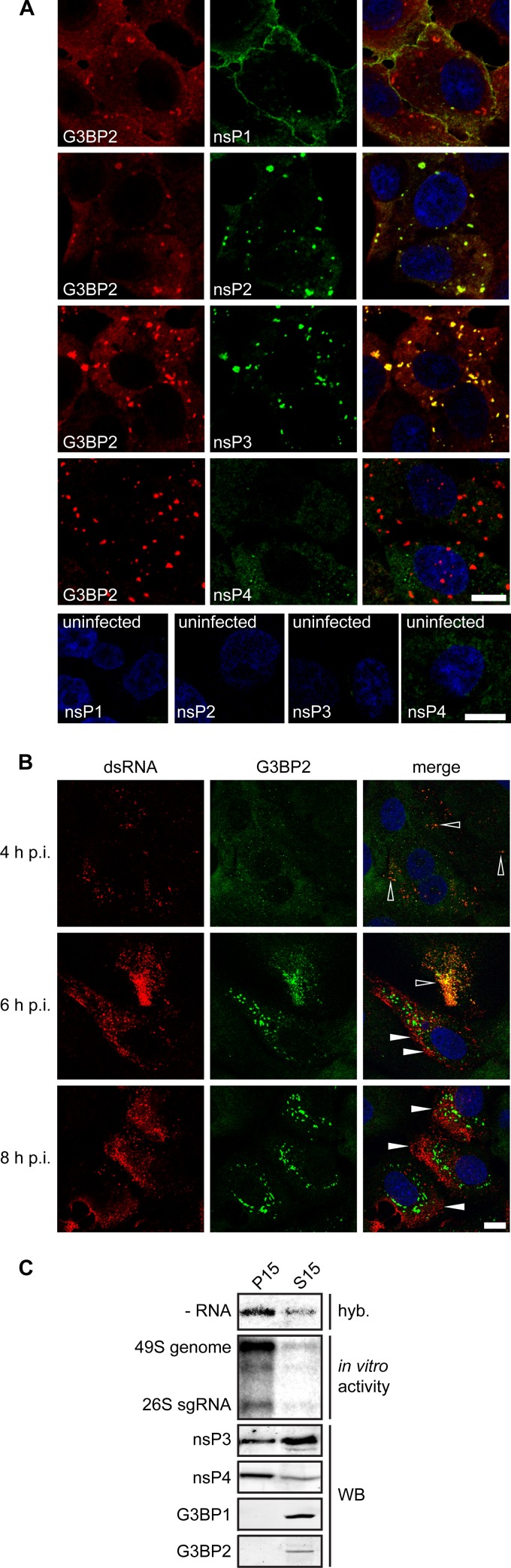
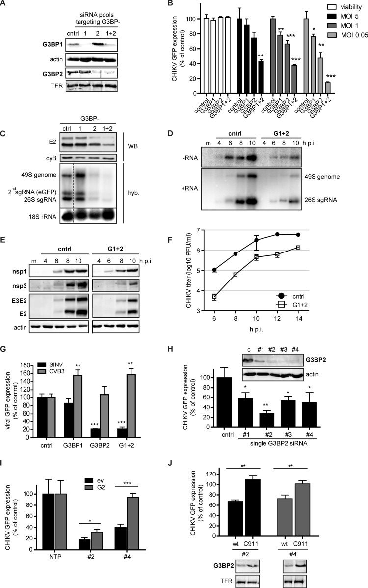
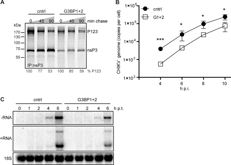
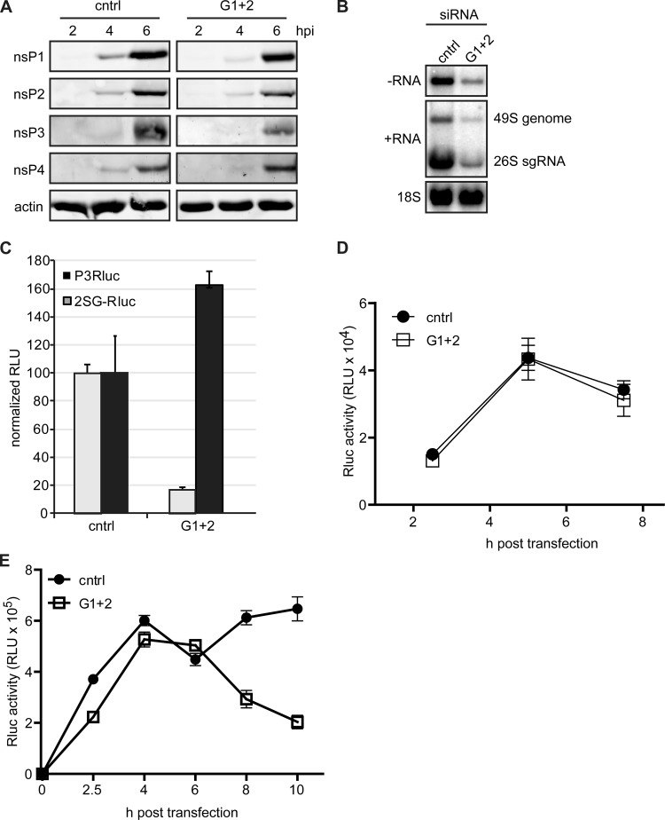
Stress granules (SGs) are protein-mRNA aggregates that are formed in response to environmental stresses, resulting in translational inhibition. SGs are generally believed to play an antiviral role and are manipulated by many viruses, including various alphaviruses. GTPase-activating protein (SH3 domain)-binding protein 1 (G3BP1) is a key component and commonly used marker of SGs. Its homolog G3BP2 is a less extensively studied SG component. Here, we demonstrate that Chikungunya virus (CHIKV) infection induces cytoplasmic G3BP1- and G3BP2-containing granules that differ from bona fide SGs in terms of morphology, composition, and behavior. For several Old World alphaviruses it has been shown that nonstructural protein 3 (nsP3) interacts with G3BPs, presumably to inhibit SG formation, and we have confirmed this interaction in CHIKV-infected cells. Surprisingly, CHIKV also relied on G3BPs for efficient replication, as simultaneous depletion of G3BP1 and G3BP2 reduced viral RNA levels, CHIKV protein expression, and viral progeny titers. The G3BPs colocalized with CHIKV nsP2 and nsP3 in cytoplasmic foci, but no colocalization with nsP1, nsP4, or dsRNA was observed. Furthermore, G3BPs could not be detected in a cellular fraction enriched for CHIKV replication/transcription complexes, suggesting that they are not directly involved in CHIKV RNA synthesis. Depletion of G3BPs did not affect viral entry, translation of incoming genomes, or nonstructural polyprotein processing but resulted in severely reduced levels of negative-stranded (and consequently also positive-stranded) RNA. This suggests a role for the G3BPs in the switch from translation to genome amplification, although the exact mechanism by which they act remains to be explored.

Importance: Chikungunya virus (CHIKV) causes a severe polyarthritis that has affected millions of people since its reemergence in 2004. The lack of approved vaccines or therapeutic options and the ongoing explosive outbreak in the Caribbean underline the importance of better understanding CHIKV replication. Stress granules (SGs) are cytoplasmic protein-mRNA aggregates formed in response to various stresses, including viral infection. The RNA-binding proteins G3BP1 and G3BP2 are essential SG components. SG formation and the resulting translational inhibition are generally considered an antiviral response, and many viruses manipulate or block this process. Late in infection, we and others have observed CHIKV nonstructural protein 3 in cytoplasmic G3BP1- and G3BP2-containing granules. These virally induced foci differed from true SGs and did not appear to represent replication complexes. Surprisingly, we found that G3BP1 and G3BP2 were also needed for efficient CHIKV replication, likely by facilitating the switch from translation to genome amplification early in infection.


Figures

FIG 1
FIG 1
Induction of G3BP-containing foci by CHIKV replication. (A) Vero E6 cells were infected with CHIKV (MOI, 5), fixed at the indicated time points postinfection, and immunostained for G3BP2 and CHIKV. A longer exposure of mock-infected cells is shown to visualize diffuse G3BP2 staining. (B) CHIKV-induced granules (MOI, 5; 8 h p.i.) were costained for G3BP1 and G3BP2. (C) Vero E6 cells were infected with CHIKV (MOI, 5; analyzed at 8 h p.i.) or treated with arsenite (0.5 mM for 1 h) to induce SG formation and subsequently incubated with CHX (100 μg/ml for 30 min), followed by immunostaining for G3BP1. (D) G3BP2 immunostaining of Vero cells that were infected with CHIKV ITA07-RA1 or LR2006-OPY1-nsP4/FLAG (MOI, 5) and fixed at 6 h p.i. Scale bar: 10 μm.
FIG 2
FIG 2
Composition of arsenite-induced SGs and CHIKV-induced granules. Vero E6 cells were treated with arsenite or infected with CHIKV-GFP (MOI, 5; fixed at 8 h p.i.). After fixation, the localization of the SG markers indicated to the left of each row was analyzed by immunofluorescence microscopy. The rightmost column shows the overlays of the signals of the SG marker and eGFP. Scale bar: 10 μm.
FIG 3
FIG 3
Levels of stress granule proteins during CHIKV infection. 293/ACE2 cells were infected with CHIKV (MOI, 5), and whole-cell lysates were prepared at the indicated time points postinfection. The expression level of the indicated proteins was determined by Western blotting. Mock-infected cells (m) were included as a negative control, and actin was used as a loading control.
FIG 4
FIG 4
Localization of CHIKV nonstructural proteins, dsRNA, and G3BP. (A) Vero cells were infected with a CHIKV encoding nsP4-FLAG (LR2006-OPY1- nsP4/FLAG) at an MOI of 5, fixed at 6 h p.i., and stained for G3BP2, nsP1, nsP2, nsP3, and nsP4/FLAG. The bottom part of panel A shows the staining of uninfected cells, with the antibodies indicated in each frame. (B) Vero E6 cells were infected with CHIKV (MOI, 5), fixed at the indicated time points p.i., and stained for G3BP2 and dsRNA. Open arrowheads indicate colocalization of dsRNA with G3BP2, whereas closed arrowheads indicate some examples of nonoverlapping signals. Scale bar: 10 μm. (C) Distribution of G3BPs and CHIKV nsPs between the “heavy membrane” fraction P15 and “cytoplasmic” fraction S15. Vero E6 cells were infected with CHIKV (MOI, 5) and then harvested, lysed, and subjected to subcellular fractionation at 6 h p.i. The presence of CHIKV negative-stranded RNA was determined by hybridization with a specific probe (hyb). The RNA synthesizing activity was assessed with an in vitro assay in which the incorporation of [32P]CTP into CHIKV RNA was determined (31). The levels of CHIKV nsP3, nsP4, G3BP1, and G3BP2 in the P15 and S15 fractions were determined by Western blotting (WB).
FIG 5
FIG 5
Effect of siRNA-mediated depletion of G3BP1 and G3BP2 on CHIKV replication. (A) Western blot analysis of the protein levels of G3BP1 and G3BP2 in 293/ACE2 cells transfected with control siRNAs or those targeting G3BP1, G3BP2, or both. The transferrin receptor (TFR) and actin were used as loading controls. (B) Cell viability and CHIKV-driven eGFP expression in cells that were depleted of G3BP1 and G3BP2 and subsequently infected with reporter virus CHIKV LS3-GFP at an MOI of 5 (black bars), 1 (dark gray bars), or 0.05 (light gray bars). Cell viability was determined at 48 h p.t. (white bars), and eGFP expression was quantified at 16, 20, or 24 h p.i., depending on the MOI used. (C) G3BP-depleted, CHIKV-infected cells (MOI, 0.05) were analyzed for E2 expression (by Western blotting using cyclophilin B as a loading control) and positive-strand RNA levels (by in-gel hybridization [hyb]) at 24 h p.i. (D) In-gel hybridization analysis of CHIKV RNA levels with probes specific for negative-strand (−RNA) or positive-strand (+RNA) RNA in G3BP-depleted 293/ACE2 cells. The cells were infected with CHIKV (MOI, 5) 48 h after siRNA transfection, and total RNA was isolated at the indicated time points. cntrl, control. (E) Western blot analysis of CHIKV protein expression levels after G3BP depletion. 293/ACE2 cells were transfected with control or G3BP-specific siRNAs and 48 h later infected with CHIKV (MOI, 5). Cell lysates for Western blot analysis were harvested at the indicated time points and analyzed for the viral proteins indicated, using actin as a loading control. (F) Infectious progeny titers of CHIKV-infected cells (MOI, 5) that were transfected with control or G3BP-specific siRNAs. (G) G3BP-depleted 293/ACE2 cells were infected with SINV-GFP or CVB3-GFP (MOI, 1) and fixed at 16 or 10 h p.i., respectively. The level of GFP expressed by the viruses was normalized to infected cells transfected with control siRNAs. (H) 293/ACE2 cells were transfected with 50 nM individual siRNA duplexes (deconvoluted pool) and infected with CHIKV-GFP (MOI, 1) 2 days later, followed by quantification of GFP expression at 20 h p.i. The (remaining) level of G3BP2 expression was determined by Western blotting. (I) Analysis of GFP expressed by CHIKV in cells depleted for G3BP2 with siRNA 2 or 4 and cotransfected with a plasmid encoding siRNA-resistant G3BP2 (G2) or an empty vector (ev). (J) Cells were transfected with C911 mutant siRNAs (C911) or the corresponding G3BP2-targeting siRNAs (wt), followed 24 h later by infection with CHIKV-GFP (MOI, 1) and quantification of GFP expression at 20 h p.i. GFP levels were normalized to cells transfected with a nontargeting control siRNA (100%). G3BP2 knockdown efficiency at 24 h p.t. was determined by Western blotting.
FIG 6
FIG 6
Effect of G3BP depletion on CHIKV nonstructural polyprotein processing, rate of RNA synthesis, and entry. 293/ACE2 cells were transfected with siRNAs targeting the G3BPs and 48 h later infected with CHIKV (MOI, 5) or transfected with in vitro-transcribed CHIKV genomic RNA. (A) Cells were metabolically labeled at 5 h p.i. and chased for 0, 45, or 90 min before lysis and immunoprecipitation. (B) Total RNA from infected cells was isolated at the indicated time points, and CHIKV genome copy numbers were determined using qRT-PCR. (C) G3BP-depleted cells were transfected with 1 μg of viral RNA (in vitro transcript) and harvested at the indicated time points. CHIKV RNA was analyzed using in-gel hybridization using probes specific for negative- or positive-strand RNA. The 18S rRNA was probed as a loading control.
FIG 7
FIG 7
Effect of G3BP depletion on translation of genomic RNA and early negative-strand RNA synthesis. (A) Cells transfected with control (cntrl) or G3BP1- and G3BP2-targeting (G1 + 2) siRNAs were infected with CHIKV at an MOI of 50 and harvested at the indicated time points, after which viral protein levels were analyzed by Western blotting. (B) siRNA-treated cells were infected with CHIKV (MOI, 50), and viral RNA was isolated at 5 h p.i. and analyzed by in-gel hybridization. The 18S rRNA was probed as a loading control. (C) G3BP-depleted cells were infected with reporter viruses that express Renilla luciferase either fused to nsP3 (P3Rluc) or from a duplicated subgenomic promoter (2SG-Rluc) at an MOI of 5. Luciferase activity was determined at 8 h p.i. and normalized to the activity in cells transfected with control siRNAs. (D) Replication-deficient CHIKV-P3Rluc RNA was transfected into control and G3BP-depleted cells and luciferase activity was assessed at the indicated time points. (E) Replication-competent CHIKV-P3Rluc RNA was transfected into control and G3BP-depleted cells, followed by measurement of luciferase activity at the indicated time points.

Similar articles

Cited by

References

    1. White JP, Lloyd RE. 2012. Regulation of stress granules in virus systems. Trends Microbiol 20:175–183. doi:10.1016/j.tim.2012.02.001. - DOI - PMC - PubMed
    1. Tsai W-C, Lloyd RE. 2014. Cytoplasmic RNA granules and viral infection. Annu Rev Virol 1:147–170. doi:10.1146/annurev-virology-031413-085505. - DOI - PMC - PubMed
    1. Kedersha N, Anderson P. 2002. Stress granules: sites of mRNA triage that regulate mRNA stability and translatability. Biochem Soc Trans 30:963–969. doi:10.1152/ajpcell.00314.2002. - DOI - PubMed
    1. Kimball SR, Horetsky RL, Ron D, Jefferson LS, Harding HP. 2003. Mammalian stress granules represent sites of accumulation of stalled translation initiation complexes. Am J Physiol Cell Physiol 284:C273–C284. doi:10.1152/ajpcell.00314.2002. - DOI - PubMed
    1. Tourrière H, Chebli K, Zekri L, Courselaud B, Blanchard JM, Bertrand E, Tazi J. 2003. The RasGAP-associated endoribonuclease G3BP assembles stress granules. J Cell Biol 160:823–831. doi:10.1083/jcb.200212128. - DOI - PMC - PubMed

Publication types

MeSH terms