. 2015 Apr;89(8):4504-16.
doi: 10.1128/JVI.00057-15. Epub 2015 Feb 4.

Influenza hemagglutinin (HA) stem region mutations that stabilize or destabilize the structure of multiple HA subtypes

Affiliations

Influenza hemagglutinin (HA) stem region mutations that stabilize or destabilize the structure of multiple HA subtypes

Lauren Byrd-Leotis et al. J Virol. 2015 Apr.

Abstract

Influenza A viruses enter host cells through endosomes, where acidification induces irreversible conformational changes of the viral hemagglutinin (HA) that drive the membrane fusion process. The prefusion conformation of the HA is metastable, and the pH of fusion can vary significantly among HA strains and subtypes. Furthermore, an accumulating body of evidence implicates HA stability properties as partial determinants of influenza host range, transmission phenotype, and pathogenic potential. Although previous studies have identified HA mutations that can affect HA stability, these have been limited to a small selection of HA strains and subtypes. Here we report a mutational analysis of HA stability utilizing a panel of expressed HAs representing a broad range of HA subtypes and strains, including avian representatives across the phylogenetic spectrum and several human strains. We focused on two highly conserved residues in the HA stem region: HA2 position 58, located at the membrane distal tip of the short helix of the hairpin loop structure, and HA2 position 112, located in the long helix in proximity to the fusion peptide. We demonstrate that a K58I mutation confers an acid-stable phenotype for nearly all HAs examined, whereas a D112G mutation consistently leads to elevated fusion pH. The results enhance our understanding of HA stability across multiple subtypes and provide an additional tool for risk assessment for circulating strains that may have other hallmarks of human adaptation. Furthermore, the K58I mutants, in particular, may be of interest for potential use in the development of vaccines with improved stability profiles.

Importance: The influenza A hemagglutinin glycoprotein (HA) mediates the receptor binding and membrane fusion functions that are essential for virus entry into host cells. While receptor binding has long been recognized for its role in host species specificity and transmission, membrane fusion and associated properties of HA stability have only recently been appreciated as potential determinants. We show here that mutations can be introduced at highly conserved positions to stabilize or destabilize the HA structure of multiple HA subtypes, expanding our knowledge base for this important phenotype. The practical implications of these findings extend to the field of vaccine design, since the HA mutations characterized here could potentially be utilized across a broad spectrum of influenza virus subtypes to improve the stability of vaccine strains or components.


Figures

FIG 1
FIG 1
(A) The H3 subtype monomer is shown in the central panel with HA1 depicted in blue and HA2 depicted in red. The structural locations of HA2 K58 and HA2 D112 are indicated. In the upper left region of the figure, the head domains corresponding shaded portion of the monomer are shown, with the H3 head domain (blue) superimposed on the H1 (red). The depiction indicates the higher location relative to HA stems for the H1 subtype, and the “twist” in rotational symmetry. The lower left panel shows the HA2 hairpin structures of representatives of four of the five clades superimposed on one another and demonstrates that all are very similar in the long helix domain, but vary in structure for the connecting peptide and top of the short helix (color-coded to match panel B). The bottom right panels correspond to the lower shaded region of the monomer structure and highlight both highly conserved residues and those that segregate in group-specific fashion. HA2 K51, D109, and D112 (top to bottom) are highly conserved, whereas HA2 R/H106, E/Q105, H/T111, and HA1 Y/H17 are conserved within groups. FP indicates the HA2 N-terminal fusion peptide domain, and the spheres denote the HA2 N-terminal glycine residue. (B) This panel shows a phylogenetic tree of the 16 subtypes that circulate in avian species, with the subtypes belonging to group 1 and group 2 indicated and the five clades color-coded to designate those subtypes more closely related by sequence and structure.
FIG 2
FIG 2
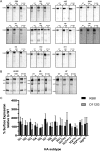
Surface expression of WT and mutant HAs. Radiolabeled, cell surface proteins were biotinylated and precipitated with an HA-specific antibody and then streptavidin where indicated (SE) and resolved by SDS-PAGE. Lanes are denoted “T” for total HA and “SE” for surface expressed. (A) Gel images for human and avian subtypes are shown. (B) Gel images for H5VN, H5, and H7 are shown. These HAs are expressed in both cleaved and uncleaved forms due to endogenous proteases. (C) Quantitation of the surface expression of mutant HA relative to WT HA. For each WT and mutant HA, the intensity of the bands corresponding to HA0 + streptavidin and HA0 − streptavidin were normalized based on methionine content. The percent surface expression was determined using the following equation (cell surface HA/total HA) × 100, and then the percent relative to wild-type surface expression was calculated as [(% surface expression mutant HA/% surface expression WT HA) × 100]. Error bars represent the standard deviation of three independent experiments.
FIG 3
FIG 3
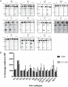
Analysis of proteolytic cleavage of WT and mutant HAs. Radiolabeled, HA-expressing cells were treated with 5 μg of TPCK-trypsin/ml, where indicated. Total cellular HA protein was precipitated from the lysate with a HA-specific antibody and resolved by SDS-PAGE. (A) Gel images for human and avian subtypes are shown. (B) Quantitation of trypsin-mediated cleavage of mutant HA relative to WT HA. For each WT and mutant HA, the intensity of the bands corresponding to HA0 and HA2 were normalized based on methionine content. For the H5VN, H5, and H7 subtypes, the cleavage values were determined from the total HA lanes of the surface expression gels shown in Fig. 2B. Percent Cleavage was determined by using the equation [HA2/(HA2 + HA0)] × 100%, and the percent cleavage relative to wild-type was calculated as [(% cleavage mutant HA/% cleavage of WT) × 100]. Error bars represent the standard deviations of three independent experiments.
FIG 4
FIG 4
Fusion pH as detected by syncytium formation. Photomicrographs of syncytium formation assays for group 1 and group 2, as well as human and avian representative HAs, are shown. HA-expressing BHK cells were treated with TPCK-trypsin (5 μg/ml) and exposed to pH adjusted PBS in 0.1 pH unit increments. Photomicrographs corresponding to the highest pH at which syncytia were observed and then 0.1 pH unit higher are shown.
FIG 5
FIG 5
Syncytium formation assay results for all HA subtypes tested. The substitution H4-K58I did not produce syncytia; the H4-D112G did mediate fusion in the syncytium assay although there was no pH difference between WT and D112G (pH 5.5). H11-K58I (pH 5.8) resulted in a higher pH of fusion that the WT H11 (pH 5.6). H5VN-D112G did mediate fusion; however, the pH of fusion was not higher than that of WT (5.6).
FIG 6
FIG 6
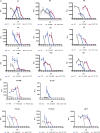
Fusion pH as detected by luciferase reporter gene assay. HA-expressing Vero cells transfected with T7-luciferase plasmid were treated with TPCK-trypsin (5 μg/ml), followed by soybean trypsin inhibitor (20 μg/ml), and then overlaid with BSR-T7/5 target cells that constitutively express T7 RNA polymerase. The mixed cell population was exposed to pH-adjusted PBS in 0.2 pH unit increments and incubated for 6 h at 37°C to allow for cell-to-cell fusion and content mixing, leading to the expression of firefly luciferase to occur. Luminescence was measured as an indicator of membrane fusion in cell lysates. The graphs show the background-adjusted luminescence. Error bars represent the standard deviations of triplicate experiments. (A) Avian origin HA subtypes. (B) Human origin HA subtypes. No fusion was detected for the H1GA-D112G HA.

Similar articles

Cited by

References

    1. Webster RG, Bean WJ, Gorman OT, Chambers TM, Kawaoka Y. 1992. Evolution and ecology of influenza A viruses. Microbiol Rev 56:152–179. - PMC - PubMed
    1. Herfst S, Imai M, Kawaoka Y, Fouchier RA. 2014. Avian influenza virus transmission to mammals. Curr Top Microbiol Immunol 385:137–155. doi:10.1007/82_2014_387. - DOI - PubMed
    1. de Jong JC, Claas EC, Osterhaus AD, Webster RG, Lim WL. 1997. A pandemic warning? Nature 389:554. doi:10.1038/39218. - DOI - PMC - PubMed
    1. Hien TT, de Jong M, Farrar J. 2004. Avian influenza: a challenge to global health care structures. N Engl J Med 351:2363–2365. doi:10.1056/NEJMp048267. - DOI - PubMed
    1. Gao R, Cao B, Hu Y, Feng Z, Wang D, Hu W, Chen J, Jie Z, Qiu H, Xu K, Xu X, Lu H, Zhu W, Gao Z, Xiang N, Shen Y, He Z, Gu Y, Zhang Z, Yang Y, Zhao X, Zhou L, Li X, Zou S, Zhang Y, Li X, Yang L, Guo J, Dong J, Li Q, Dong L, Zhu Y, Bai T, Wang S, Hao P, Yang W, Zhang Y, Han J, Yu H, Li D, Gao GF, Wu G, Wang Y, Yuan Z, Shu Y. 2013. Human infection with a novel avian-origin influenza A (H7N9) virus. N Engl J Med 368:1888–1897. doi:10.1056/NEJMoa1304459. - DOI - PubMed

Publication types