

Cannabinoid receptor CB2 is involved in tetrahydrocannabinol-induced anti-inflammation against lipopolysaccharide in MG-63 cells
- PMID: 25653478
- PMCID: PMC4310496
- DOI: 10.1155/2015/362126
Cannabinoid receptor CB2 is involved in tetrahydrocannabinol-induced anti-inflammation against lipopolysaccharide in MG-63 cells
Abstract
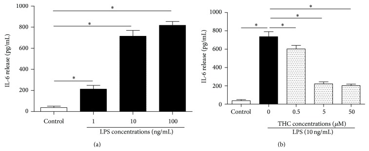
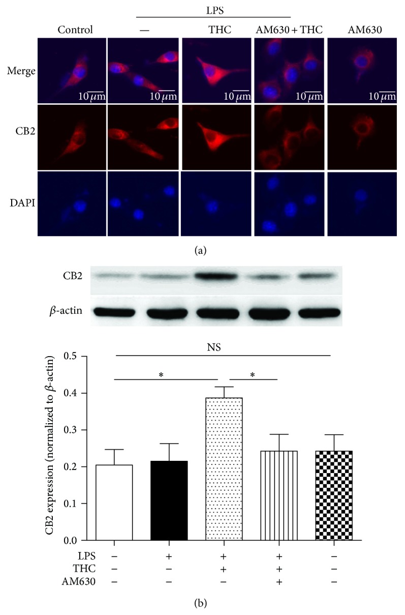
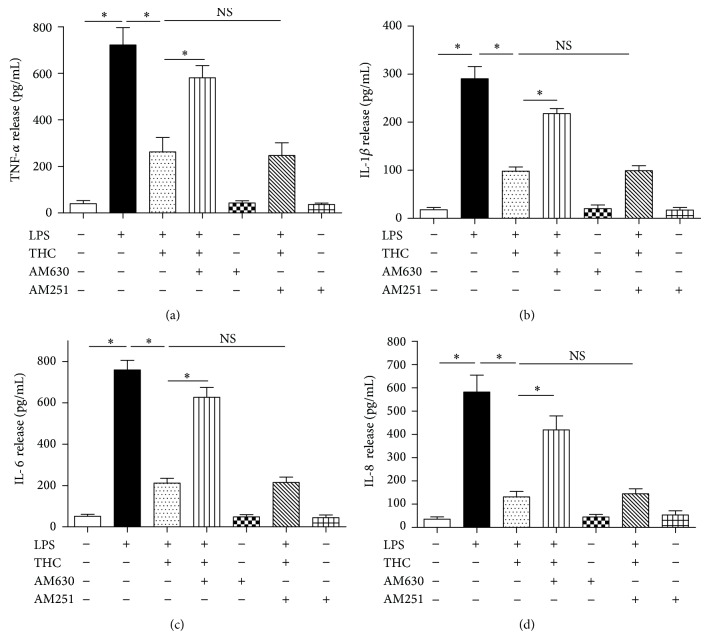
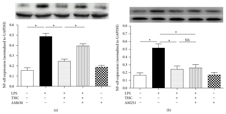
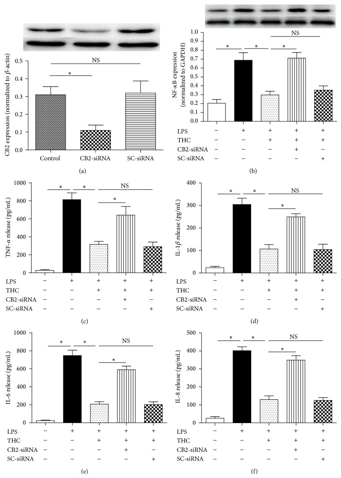
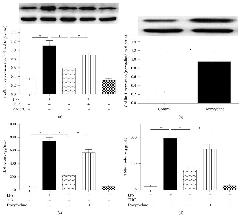
Cannabinoid Δ9-tetrahydrocannabinol (THC) is effective in treating osteoarthritis (OA), and the mechanism, however, is still elusive. Activation of cannabinoid receptor CB2 reduces inflammation; whether the activation CB2 is involved in THC-induced therapeutic action for OA is still unknown. Cofilin-1 is a cytoskeleton protein, participating in the inflammation of OA. In this study, MG-63 cells, an osteosarcoma cell-line, were exposed to lipopolysaccharide (LPS) to mimic the inflammation of OA. We hypothesized that the activation of CB2 is involved in THC-induced anti-inflammation in the MG-63 cells exposed to LPS, and the anti-inflammation is mediated by cofilin-1. We found that THC suppressed the release of proinflammatory factors, including tumor necrosis factor α (TNF-α), interleukin- (IL-) 1β, IL-6, and IL-8, decreased nuclear factor-κB (NF-κB) expression, and inhibited the upregulation of cofilin-1 protein in the LPS-stimulated MG-63 cells. However, administration of CB2 receptor antagonist or the CB2-siRNA, not CB1 antagonist AM251, partially abolished the THC-induced anti-inflammatory effects above. In addition, overexpression of cofilin-1 significantly reversed the THC-induced anti-inflammatory effects in MG-63 cells. These results suggested that CB2 is involved in the THC-induced anti-inflammation in LPS-stimulated MG-63 cells, and the anti-inflammation may be mediated by cofilin-1.
Figures
Similar articles
-
Δ9-Tetrahydrocannabinol reverses TNFα-induced increase in airway epithelial cell permeability through CB2 receptors.Biochem Pharmacol. 2016 Nov 15;120:63-71. doi: 10.1016/j.bcp.2016.09.008. Epub 2016 Sep 15. Biochem Pharmacol. 2016. PMID: 27641813
-
Effects of targeted deletion of cannabinoid receptors CB1 and CB2 on immune competence and sensitivity to immune modulation by Delta9-tetrahydrocannabinol.J Leukoc Biol. 2008 Dec;84(6):1574-84. doi: 10.1189/jlb.0508282. Epub 2008 Sep 12. J Leukoc Biol. 2008. PMID: 18791168 Free PMC article.
-
Cannabinoids Delta(9)-tetrahydrocannabinol and cannabidiol differentially inhibit the lipopolysaccharide-activated NF-kappaB and interferon-beta/STAT proinflammatory pathways in BV-2 microglial cells.J Biol Chem. 2010 Jan 15;285(3):1616-26. doi: 10.1074/jbc.M109.069294. Epub 2009 Nov 12. J Biol Chem. 2010. PMID: 19910459 Free PMC article.
-
Up-regulation of immunomodulatory effects of mouse bone-marrow derived mesenchymal stem cells by tetrahydrocannabinol pre-treatment involving cannabinoid receptor CB2.Oncotarget. 2016 Feb 9;7(6):6436-47. doi: 10.18632/oncotarget.7042. Oncotarget. 2016. PMID: 26824325 Free PMC article.
-
Molecular Mechanisms for the Inflammation-Resolving Actions of Lenabasum.Mol Pharmacol. 2021 Feb;99(2):125-132. doi: 10.1124/molpharm.120.000083. Epub 2020 Nov 25. Mol Pharmacol. 2021. PMID: 33239333 Review.
Cited by
-
G Protein-Coupled Receptors in Osteoarthritis: A Novel Perspective on Pathogenesis and Treatment.Front Cell Dev Biol. 2021 Oct 20;9:758220. doi: 10.3389/fcell.2021.758220. eCollection 2021. Front Cell Dev Biol. 2021. PMID: 34746150 Free PMC article. Review.
-
Osteosarcoma in Children: Not Only Chemotherapy.Pharmaceuticals (Basel). 2021 Sep 13;14(9):923. doi: 10.3390/ph14090923. Pharmaceuticals (Basel). 2021. PMID: 34577623 Free PMC article. Review.
-
The Endocannabinoid/Endovanilloid System in Bone: From Osteoporosis to Osteosarcoma.Int J Mol Sci. 2019 Apr 18;20(8):1919. doi: 10.3390/ijms20081919. Int J Mol Sci. 2019. PMID: 31003519 Free PMC article. Review.
-
Development of a Cannabinoid-Based Photoaffinity Probe to Determine the Δ8/9-Tetrahydrocannabinol Protein Interaction Landscape in Neuroblastoma Cells.Cannabis Cannabinoid Res. 2018 Jul 1;3(1):136-151. doi: 10.1089/can.2018.0003. eCollection 2018. Cannabis Cannabinoid Res. 2018. PMID: 29992186 Free PMC article.
-
Daily Cannabis Use is Associated With Lower CNS Inflammation in People With HIV.J Int Neuropsychol Soc. 2021 Jul;27(6):661-672. doi: 10.1017/S1355617720001447. J Int Neuropsychol Soc. 2021. PMID: 34261550 Free PMC article.
References
-
- Liu X., Xu Y., Chen S., et al. Rescue of proinflammatory cytokine-inhibited chondrogenesis by the antiarthritic effect of melatonin in synovium mesenchymal stem cells via suppression of reactive oxygen species and matrix metalloproteinases. Free Radical Biology and Medicine. 2014;68:234–246. doi: 10.1016/j.freeradbiomed.2013.12.012. - DOI - PubMed
-
- Karsdal M. A., Bay-Jensen A. C., Lories R. J., et al. The coupling of bone and cartilage turnover in osteoarthritis: opportunities for bone antiresorptives and anabolics as potential treatments? Annals of the Rheumatic Diseases. 2014;73(2):336–348. doi: 10.1136/annrheumdis-2013-204111. - DOI - PubMed
-
- Tu Y., Xue H., Francis W., et al. Lactoferrin inhibits dexamethasone-induced chondrocyte impairment from osteoarthritic cartilage through up-regulation of extracellular signal-regulated kinase 1/2 and suppression of FASL, FAS, and Caspase 3. Biochemical and Biophysical Research Communications. 2013;441(1):249–255. doi: 10.1016/j.bbrc.2013.10.047. - DOI - PubMed
Publication types
MeSH terms
Substances
LinkOut - more resources
Full Text Sources
Other Literature Sources
