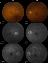
Topical difluprednate for the treatment of Harada's disease
- PMID: 25653498
- PMCID: PMC4310279
- DOI: 10.2147/OPTH.S72955
Topical difluprednate for the treatment of Harada's disease
Abstract
Purpose: To describe the use of topical difluprednate for treatment of patients who presented with Harada's disease.
Methods: Retrospective case series of patients managed with topical difluprednate alone at the onset of the diagnosis. The patients were followed with optical coherence tomography (OCT) and fluorescein angiography.
Results: In our three cases, complete resolution of the exudative detachments with improvement in visual acuity was achieved in each case. Central macular thickness was reduced by a mean of 365±222 μm. Initial loading dose was one drop every hour while awake, followed by a variable tapering regimen. Leakage on fluorescein angiography and exudative detachments on OCT both responded to treatment with difluprednate. In two of the three cases, the patients recovered vision to 20/20, and the third case recovered to 20/25. Steroid-induced glaucoma was observed and managed with one to two glaucoma drops as needed.
Conclusion: Difluprednate is effective for managing ocular manifestations of Harada's disease. Further studies of this drug for the management of noninfectious posterior uveitis are warranted.
Keywords: noninfectious posterior uveitis; serous retinal detachment; steroid.
Figures


Similar articles
-
Strong topical steroid, NSAID, and carbonic anhydrase inhibitor cocktail for treatment of cystoid macular edema.Int Med Case Rep J. 2015 Dec 1;8:305-12. doi: 10.2147/IMCRJ.S92794. eCollection 2015. Int Med Case Rep J. 2015. PMID: 26664246 Free PMC article.
-
RESOLUTION OF NONINFECTIOUS UVEITIC CYSTOID MACULAR EDEMA WITH TOPICAL DIFLUPREDNATE.Retina. 2017 May;37(5):844-850. doi: 10.1097/IAE.0000000000001243. Retina. 2017. PMID: 27529841
-
Efficacy and potential complications of difluprednate use for pediatric uveitis.Am J Ophthalmol. 2012 May;153(5):932-8. doi: 10.1016/j.ajo.2011.10.008. Epub 2012 Jan 20. Am J Ophthalmol. 2012. PMID: 22265149
-
Uveitic macular oedema: correlation between optical coherence tomography patterns with visual acuity and fluorescein angiography.Br J Ophthalmol. 2008 Jul;92(7):922-7. doi: 10.1136/bjo.2007.136846. Br J Ophthalmol. 2008. PMID: 18577643
-
[Measurement of retinal circulation in uveitis of Behçet's and Harada's disease].Nippon Ganka Gakkai Zasshi. 1989 Jun;93(6):705-13. Nippon Ganka Gakkai Zasshi. 1989. PMID: 2816579 Japanese.
Cited by
-
Topical difluprednate for the treatment of retinal vasculitis associated with birdshot chorioretinitis.Am J Ophthalmol Case Rep. 2016 Apr 29;3:22-24. doi: 10.1016/j.ajoc.2016.04.009. eCollection 2016 Oct. Am J Ophthalmol Case Rep. 2016. PMID: 29503901 Free PMC article.
-
Risk of Elevated Intraocular Pressure With Difluprednate in Patients With Non-Infectious Uveitis.Am J Ophthalmol. 2022 Aug;240:232-238. doi: 10.1016/j.ajo.2022.03.026. Epub 2022 Apr 2. Am J Ophthalmol. 2022. PMID: 35381204 Free PMC article.
-
Topical Anti-Inflammatory Agents for Non-Infectious Uveitis: Current Treatment and Perspectives.J Inflamm Res. 2022 Nov 28;15:6439-6451. doi: 10.2147/JIR.S288294. eCollection 2022. J Inflamm Res. 2022. PMID: 36467992 Free PMC article. Review.
-
Strong topical steroid, NSAID, and carbonic anhydrase inhibitor cocktail for treatment of cystoid macular edema.Int Med Case Rep J. 2015 Dec 1;8:305-12. doi: 10.2147/IMCRJ.S92794. eCollection 2015. Int Med Case Rep J. 2015. PMID: 26664246 Free PMC article.
-
Vogt-Koyanagi-Harada disease: review of a rare autoimmune disease targeting antigens of melanocytes.Orphanet J Rare Dis. 2016 Mar 24;11:29. doi: 10.1186/s13023-016-0412-4. Orphanet J Rare Dis. 2016. PMID: 27008848 Free PMC article. Review.
References
-
- Andreoli CM, Foster CS. Vogt-Koyanagi-Harada disease. Int Ophthalmol Clin. 2006;46(2):111–122. - PubMed
-
- Read RW, Holland GN, Rao NA, et al. Revised diagnostic criteria for Vogt-Koyanagi-Harada disease: report of an international committee on nomenclature. Am J Ophthalmol. 2001;131:647–652. - PubMed
-
- Weisz JM, Holland GN, Roer LN, et al. Association between Vogt-Koyanagi-Harada syndrome and HLA-DR1 and -DR4 in Hispanic patients living in Southern California. Ophthalmology. 1995;102(7):1012–1015. - PubMed
-
- Davis JL, Mittal KK, Freidlin V, et al. HLA associations and ancestry in Vogt-Koyanagi-Harada disease and sympathetic ophthalmia. Ophthalmology. 1990;97(9):1137–1142. - PubMed
LinkOut - more resources
Full Text Sources

