

Integration of modeling with experimental and clinical findings synthesizes and refines the central role of inositol 1,4,5-trisphosphate receptor 1 in spinocerebellar ataxia
- PMID: 25653583
- PMCID: PMC4300941
- DOI: 10.3389/fnins.2014.00453
Integration of modeling with experimental and clinical findings synthesizes and refines the central role of inositol 1,4,5-trisphosphate receptor 1 in spinocerebellar ataxia
Abstract
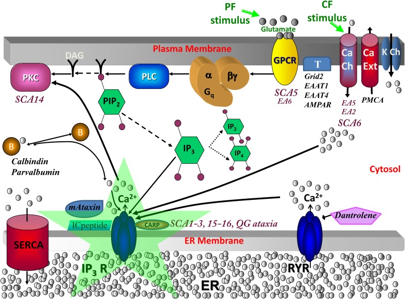
A suite of models was developed to study the role of inositol 1,4,5-trisphosphate receptor 1 (IP3R1) in spinocerebellar ataxias (SCAs). Several SCAs are linked to reduced abundance of IP3R1 or to supranormal sensitivity of the receptor to activation by its ligand inositol 1,4,5-trisphosphate (IP3). Detailed multidimensional models have been created to simulate biochemical calcium signaling and membrane electrophysiology in cerebellar Purkinje neurons. In these models, IP3R1-mediated calcium release is allowed to interact with ion channel response on the cell membrane. Experimental findings in mice and clinical observations in humans provide data input for the models. The SCA modeling suite helps interpret experimental results and provides suggestions to guide experiments. The models predict IP3R1 supersensitivity in SCA1 and compensatory mechanisms in SCA1, SCA2, and SCA3. Simulations explain the impact of calcium buffer proteins. Results show that IP3R1-mediated calcium release activates voltage-gated calcium-activated potassium channels in the plasma membrane. The SCA modeling suite unifies observations from experiments in a number of SCAs. The cadre of simulations demonstrates the central role of IP3R1.
Keywords: Purkinje; carbonic anhydrase related proteins; computational; homer; inositol 1,4,5-trisphosphate receptor 1; model; spinocerebellar ataxia; translational.
Figures
Similar articles
-
Computational analysis of calcium signaling and membrane electrophysiology in cerebellar Purkinje neurons associated with ataxia.BMC Syst Biol. 2012 Jun 15;6:70. doi: 10.1186/1752-0509-6-70. BMC Syst Biol. 2012. PMID: 22703638 Free PMC article.
-
Roles of inositol 1,4,5-trisphosphate receptors in spinocerebellar ataxias.Neurochem Int. 2016 Mar;94:1-8. doi: 10.1016/j.neuint.2016.01.007. Epub 2016 Jan 28. Neurochem Int. 2016. PMID: 26827887 Review.
-
Calcium Signaling, PKC Gamma, IP3R1 and CAR8 Link Spinocerebellar Ataxias and Purkinje Cell Dendritic Development.Curr Neuropharmacol. 2018 Jan 30;16(2):151-159. doi: 10.2174/1570159X15666170529104000. Curr Neuropharmacol. 2018. PMID: 28554312 Free PMC article. Review.
-
Aberrant IP3 receptor activities revealed by comprehensive analysis of pathological mutations causing spinocerebellar ataxia 29.Proc Natl Acad Sci U S A. 2018 Nov 27;115(48):12259-12264. doi: 10.1073/pnas.1811129115. Epub 2018 Nov 14. Proc Natl Acad Sci U S A. 2018. PMID: 30429331 Free PMC article.
-
Ca2+ signaling and spinocerebellar ataxia.Biochim Biophys Acta Mol Cell Res. 2018 Nov;1865(11 Pt B):1733-1744. doi: 10.1016/j.bbamcr.2018.05.009. Epub 2018 May 16. Biochim Biophys Acta Mol Cell Res. 2018. PMID: 29777722 Review.
Cited by
-
Modulation and Regulation of Canonical Transient Receptor Potential 3 (TRPC3) Channels.Cells. 2023 Sep 5;12(18):2215. doi: 10.3390/cells12182215. Cells. 2023. PMID: 37759438 Free PMC article. Review.
-
Lysosomal Calcium in Neurodegeneration.Messenger (Los Angel). 2016 Jun;5(1-2):56-66. doi: 10.1166/msr.2016.1055. Epub 2016 Jun 1. Messenger (Los Angel). 2016. PMID: 29082116 Free PMC article.
-
Poetic Science: Bidirectional Reflection in Science and Medicine.Perm J. 2019;23:17-177. doi: 10.7812/TPP/17-177. Epub 2019 Jul 8. Perm J. 2019. PMID: 31314715 Free PMC article.
-
Determining the Roles of Inositol Trisphosphate Receptors in Neurodegeneration: Interdisciplinary Perspectives on a Complex Topic.Mol Neurobiol. 2017 Nov;54(9):6870-6884. doi: 10.1007/s12035-016-0205-8. Epub 2016 Oct 22. Mol Neurobiol. 2017. PMID: 27771899 Review.
-
Principles for Developing Patient Avatars in Precision and Systems Medicine.Front Genet. 2016 Jan 8;6:365. doi: 10.3389/fgene.2015.00365. eCollection 2015. Front Genet. 2016. PMID: 26779255 Free PMC article. No abstract available.
References
-
- Brown S.-A., Holmes R., Loew L. M. (2012). Spatial organization and diffusion in neuronal signaling in Computational Systems Neurobiology, ed Novere N. L. (Dordrecht: Springer; ), 133–161.
Publication types
Grants and funding
LinkOut - more resources
Full Text Sources
Other Literature Sources
Research Materials