Review
. 2015 Jan 21:8:1066.
doi: 10.3389/fnhum.2014.01066. eCollection 2014.

Recent advancements in diffusion MRI for investigating cortical development after preterm birth-potential and pitfalls

Affiliations
Review

Recent advancements in diffusion MRI for investigating cortical development after preterm birth-potential and pitfalls

J Dudink et al. Front Hum Neurosci. .

Abstract

Preterm infants are born during a critical period of brain maturation, in which even subtle events can result in substantial behavioral, motor and cognitive deficits, as well as psychiatric diseases. Recent evidence shows that the main source for these devastating disabilities is not necessarily white matter (WM) damage but could also be disruptions of cortical microstructure. Animal studies showed how moderate hypoxic-ischemic conditions did not result in significant neuronal loss in the developing brain, but did cause significantly impaired dendritic growth and synapse formation alongside a disturbed development of neuronal connectivity as measured using diffusion magnetic resonance imaging (dMRI). When using more advanced acquisition settings such as high-angular resolution diffusion imaging (HARDI), more advanced reconstruction methods can be applied to investigate the cortical microstructure with higher levels of detail. Recent advances in dMRI acquisition and analysis have great potential to contribute to a better understanding of neuronal connectivity impairment in preterm birth. We will review the current understanding of abnormal preterm cortical development, novel approaches in dMRI, and the pitfalls in scanning vulnerable preterm infants.

Keywords: DTI; cortical development; cortical development and plasticity; cortical imaging technique; diffusion MRI; diffusion magnetic resonance imaging; prematurity.


Figures
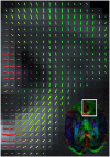
Figure 1
Figure 1
Color-coded orientation map of a preterm infant’s brain (born at 26 weeks of gestation and scanned at 30 weeks gestation at 1.5 Tesla MRI scanner) calculated from diffusion tensor imaging. Each pixel contains information about the eigenvector (Green color signifies preferential diffusion in the antero-posterior direction; Red in the latero-lateral direction; Blue in the superior-inferior direction) and extent of diffusion anisotropy (intensity of color) which summarizes the information obtained from each 3 × 3-matrix tensor. The cortex at 30 weeks gestation still has a clear radial organization (shown in close-up).
Figure 2
Figure 2
(A) High resolution diffusion-MRI of sub-cortical white matter and cortical gray matter of the adult human primary visual cortex. High angular and spatial resolution data (768 directions at 300 micrometers isotropic resolution) was used to generate orientation distribution functions (ODF’s) for each voxel. The spikes of the ODF are color-coded and represent the different directions of orientation within a single voxel. The ODF’s in the cortex are mainly orientated perpendicular to the cortical surface, and represent its radial organization, whereas underlying white matter is arranged corticofugally. (B) Close-up of the cortex in a neighboring slice, showing that the ODF’s are able to discern different layers of the cortex based on differences in microstructural characteristics between different laminae. The fractional anisotropy image is used as a backdrop image. Greyscale insets represent the anatomical GRE reference images showing cortical layers including the stria of Gennari. Adapted from Kleinnijenhuis et al. (2011).

Similar articles

Cited by

References

    1. Aggarwal M., Nauen D. W., Troncoso J. C., Mori S. (2015). Probing region-specific microstructure of human cortical areas using high angular and spatial resolution diffusion MRI. Neuroimage 105, 198–207. 10.1016/j.neuroimage.2014.10.053 - DOI - PMC - PubMed
    1. Andiman S. E., Haynes R. L., Trachtenberg F. L., Billiards S. S., Folkerth R. D., Volpe J. J., et al. . (2010). The cerebral cortex overlying periventricular leukomalacia: analysis of pyramidal neurons. Brain Pathol. 20, 803–814. 10.1111/j.1750-3639.2010.00380.x - DOI - PMC - PubMed
    1. Assaf Y., Basser P. J. (2005). Composite hindered and restricted model of diffusion (CHARMED) MR imaging of the human brain. Neuroimage 27, 48–58. 10.1016/j.neuroimage.2005.03.042 - DOI - PubMed
    1. Ball G., Boardman J. P., Aljabar P., Pandit A., Arichi T., Merchant N., et al. . (2013a). The influence of preterm birth on the developing thalamocortical connectome. Cortex 49, 1711–1721. 10.1016/j.cortex.2012.07.006 - DOI - PubMed
    1. Ball G., Srinivasan L., Aljabar P., Counsell S. J., Durighel G., Hajnal J. V., et al. . (2013b). Development of cortical microstructure in the preterm human brain. Proc. Natl. Acad. Sci. U S A 110, 9541–9546. 10.1073/pnas.1301652110 - DOI - PMC - PubMed

LinkOut - more resources