

Distributed functions of detection and discrimination of vibrotactile stimuli in the hierarchical human somatosensory system
- PMID: 25653609
- PMCID: PMC4301016
- DOI: 10.3389/fnhum.2014.01070
Distributed functions of detection and discrimination of vibrotactile stimuli in the hierarchical human somatosensory system
Abstract
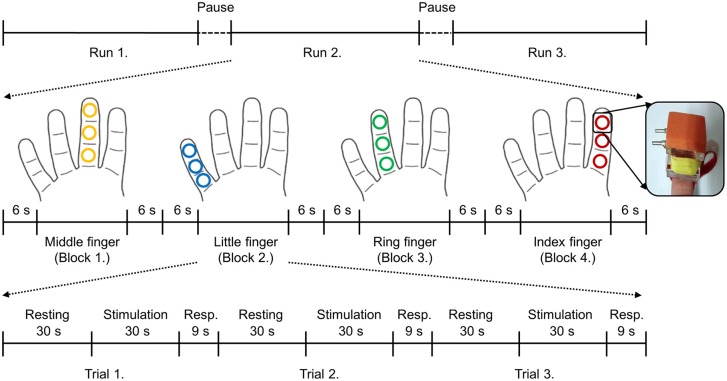
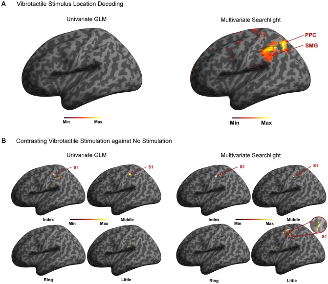
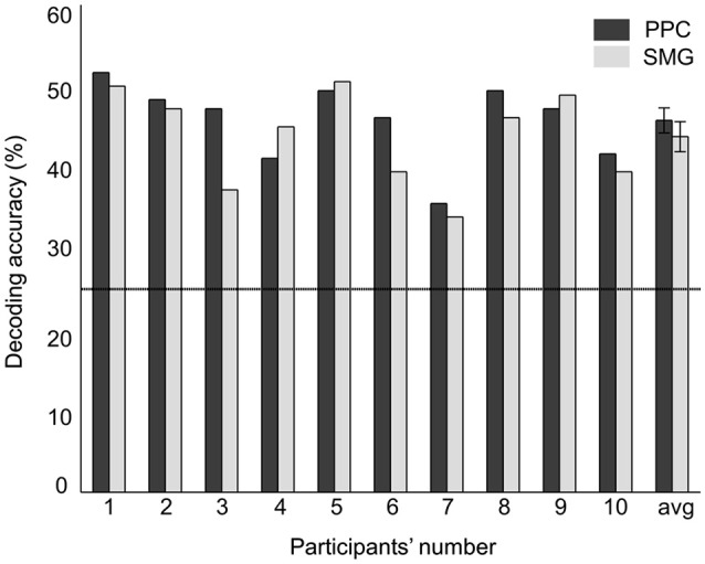
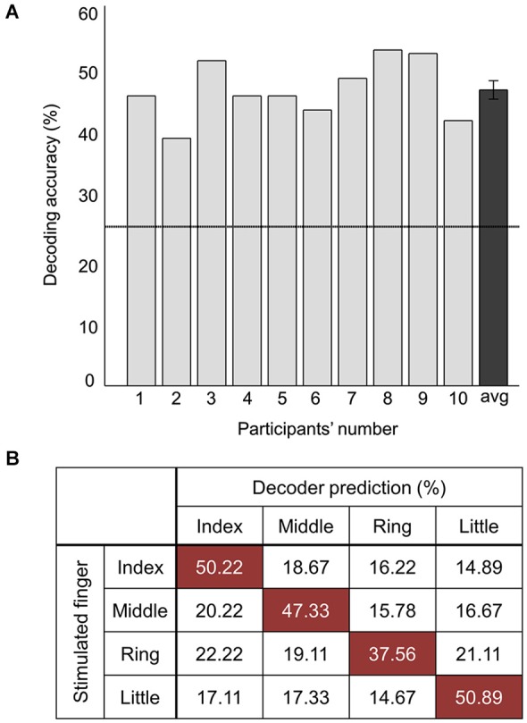
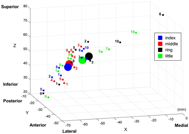
According to the hierarchical view of human somatosensory network, somatic sensory information is relayed from the thalamus to primary somatosensory cortex (S1), and then distributed to adjacent cortical regions to perform further perceptual and cognitive functions. Although a number of neuroimaging studies have examined neuronal activity correlated with tactile stimuli, comparatively less attention has been devoted toward understanding how vibrotactile stimulus information is processed in the hierarchical somatosensory cortical network. To explore the hierarchical perspective of tactile information processing, we studied two cases: (a) discrimination between the locations of finger stimulation; and (b) detection of stimulation against no stimulation on individual fingers, using both standard general linear model (GLM) and searchlight multi-voxel pattern analysis (MVPA) techniques. These two cases were studied on the same data set resulting from a passive vibrotactile stimulation experiment. Our results showed that vibrotactile stimulus locations on fingers could be discriminated from measurements of human functional magnetic resonance imaging (fMRI). In particular, it was in case (a) we observed activity in contralateral posterior parietal cortex (PPC) and supramarginal gyrus (SMG) but not in S1, while in case; (b) we found significant cortical activations in S1 but not in PPC and SMG. These discrepant observations suggest the functional specialization with regard to vibrotactile stimulus locations, especially, the hierarchical information processing in the human somatosensory cortical areas. Our findings moreover support the general understanding that S1 is the main sensory receptive area for the sense of touch, and adjacent cortical regions (i.e., PPC and SMG) are in charge of a higher level of processing and may thus contribute most for the successful classification between stimulated finger locations.
Keywords: fMRI; functional specialization; hierarchical tactile processing; somatosensory cortex; vibrotactile stimulation.
Figures
Similar articles
-
Spatial Information of Somatosensory Stimuli in the Brain: Multivariate Pattern Analysis of Functional Magnetic Resonance Imaging Data.Neural Plast. 2020 Jun 29;2020:8307580. doi: 10.1155/2020/8307580. eCollection 2020. Neural Plast. 2020. PMID: 32684924 Free PMC article.
-
Decoding pressure stimulation locations on the fingers from human neural activation patterns.Neuroreport. 2016 Nov 9;27(16):1232-6. doi: 10.1097/WNR.0000000000000683. Neuroreport. 2016. PMID: 27631540
-
Performing a vibrotactile discrimination task modulates finger representations in primary somatosensory cortex.J Neurophysiol. 2023 Oct 1;130(4):1015-1027. doi: 10.1152/jn.00428.2022. Epub 2023 Sep 6. J Neurophysiol. 2023. PMID: 37671429 Free PMC article.
-
Involvement of human primary somatosensory cortex in vibrotactile detection depends on task demand.Neuroimage. 2016 Sep;138:184-196. doi: 10.1016/j.neuroimage.2016.05.056. Epub 2016 May 24. Neuroimage. 2016. PMID: 27233148
-
Feeling for space or for time: task-dependent modulation of the cortical representation of identical vibrotactile stimuli.Neurosci Lett. 2010 Aug 16;480(2):143-7. doi: 10.1016/j.neulet.2010.06.027. Epub 2010 Jun 16. Neurosci Lett. 2010. PMID: 20561566
Cited by
-
Comparative density of CCK- and PV-GABA cells within the cortex and hippocampus.Front Neuroanat. 2015 Sep 23;9:124. doi: 10.3389/fnana.2015.00124. eCollection 2015. Front Neuroanat. 2015. PMID: 26441554 Free PMC article.
-
Human Foot Outperforms the Hand in Mechanical Pain Discrimination.eNeuro. 2024 Feb 15;11(2):ENEURO.0412-23.2024. doi: 10.1523/ENEURO.0412-23.2024. Print 2024 Feb. eNeuro. 2024. PMID: 38272674 Free PMC article.
-
Resting-state functional connectivity changes following audio-tactile speech training.Front Neurosci. 2025 Apr 29;19:1482828. doi: 10.3389/fnins.2025.1482828. eCollection 2025. Front Neurosci. 2025. PMID: 40364857 Free PMC article.
-
Shared neural representations of tactile roughness intensities by somatosensation and touch observation using an associative learning method.Sci Rep. 2019 Jan 11;9(1):77. doi: 10.1038/s41598-018-37378-w. Sci Rep. 2019. PMID: 30635598 Free PMC article.
-
Decoding spatial location of perceived pain to acupuncture needle using multivoxel pattern analysis.Mol Pain. 2019 Jan-Dec;15:1744806919877060. doi: 10.1177/1744806919877060. Mol Pain. 2019. PMID: 31469030 Free PMC article.
References
LinkOut - more resources
Full Text Sources
Other Literature Sources
Medical