Serious Complications after Self-expandable Metallic Stent Insertion in a Patient with Malignant Lymphoma
- PMID: 25653695
- PMCID: PMC4311033
- DOI: 10.4046/trd.2015.78.1.31
Serious Complications after Self-expandable Metallic Stent Insertion in a Patient with Malignant Lymphoma
Abstract
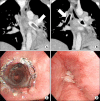
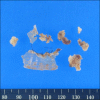
An 18-year-old woman was evaluated for a chronic productive cough and dyspnea. She was subsequently diagnosed with mediastinal non-Hodgkin lymphoma (NHL). A covered self-expandable metallic stent (SEMS) was implanted to relieve narrowing in for both main bronchi. The NHL went into complete remission after six chemotherapy cycles, but atelectasis developed in the left lower lobe 18 months after SEMS insertion. The left main bronchus was completely occluded by granulation tissue. However, the right main bronchus and intermedius bronchus were patent. Granulation tissue was observed adjacent to the SEMS. The granulation tissue and the SEMS were excised, and a silicone stent was successfully implanted using a rigid bronchoscope. SEMS is advantageous owing to its easy implantation, but there are considerable potential complications such as severe reactive granulation, stent rupture, and ventilation failure in serious cases. Therefore, SEMS should be avoided whenever possible in patients with benign airway disease. This case highlights that SEMS implantation should be avoided even in malignant airway obstruction cases if the underlying malignancy is curable.
Keywords: Airway Obstruction; Bronchoscopy; Granulation Tissue; Stents.
Conflict of interest statement
No potential conflict of interest relevant to this article was reported.
Figures


Similar articles
-
Clinical Outcomes of Complications Following Self-Expandable Metallic Stent Insertion for Benign Tracheobronchial Stenosis.Medicina (Kaunas). 2020 Jul 22;56(8):367. doi: 10.3390/medicina56080367. Medicina (Kaunas). 2020. PMID: 32708022 Free PMC article.
-
Safety and Efficacy of a Fully Covered Self-Expandable Metallic Stent in Benign Airway Stenosis.Respiration. 2017;93(6):430-435. doi: 10.1159/000472155. Epub 2017 Apr 28. Respiration. 2017. PMID: 28448981
-
Removal of self expandable metallic airway stent: A rare case report.Lung India. 2013 Jan;30(1):64-6. doi: 10.4103/0970-2113.106177. Lung India. 2013. PMID: 23661920 Free PMC article.
-
Self-expandable metallic stents in nonmalignant large airway disease.Can Respir J. 2015 Jul-Aug;22(4):235-6. doi: 10.1155/2015/246509. Can Respir J. 2015. PMID: 26252535 Free PMC article. Review.
-
Expandable stents.Chest Surg Clin N Am. 1996 May;6(2):305-28. Chest Surg Clin N Am. 1996. PMID: 8724281 Review.
Cited by
-
Rescue patient from tracheal obstruction by dislocated bronchial stent during tracheostomy surgery with readily available tools: A case report.Medicine (Baltimore). 2017 Sep;96(36):e7841. doi: 10.1097/MD.0000000000007841. Medicine (Baltimore). 2017. PMID: 28885335 Free PMC article.
-
Clinical Outcomes of Complications Following Self-Expandable Metallic Stent Insertion for Benign Tracheobronchial Stenosis.Medicina (Kaunas). 2020 Jul 22;56(8):367. doi: 10.3390/medicina56080367. Medicina (Kaunas). 2020. PMID: 32708022 Free PMC article.
-
Internal fixation of the proximal tracheal self-expandable metallic stent (SEMS): migration prevention in high risk patients.J Thorac Dis. 2020 Jun;12(6):3211-3216. doi: 10.21037/jtd-20-642. J Thorac Dis. 2020. PMID: 32642242 Free PMC article.
References
-
- Bolliger CT, Sutedja TG, Strausz J, Freitag L. Therapeutic bronchoscopy with immediate effect: laser, electrocautery, argon plasma coagulation and stents. Eur Respir J. 2006;27:1258–1271. - PubMed
-
- Casal RF. Update in airway stents. Curr Opin Pulm Med. 2010;16:321–328. - PubMed
-
- Ernst A, Feller-Kopman D, Becker HD, Mehta AC. Central airway obstruction. Am J Respir Crit Care Med. 2004;169:1278–1297. - PubMed
-
- Chin CS, Litle V, Yun J, Weiser T, Swanson SJ. Airway stents. Ann Thorac Surg. 2008;85:S792–S796. - PubMed
-
- Ryu YJ, Kim H, Yu CM, Choi JC, Kwon YS, Kwon OJ. Use of silicone stents for the management of post-tuberculosis tracheobronchial stenosis. Eur Respir J. 2006;28:1029–1035. - PubMed
LinkOut - more resources
Full Text Sources
Other Literature Sources

