

Two cases of immediate stent fracture after zotarolimus-eluting stent implantation
- PMID: 25653706
- PMCID: PMC4310982
- DOI: 10.4070/kcj.2015.45.1.67
Two cases of immediate stent fracture after zotarolimus-eluting stent implantation
Abstract
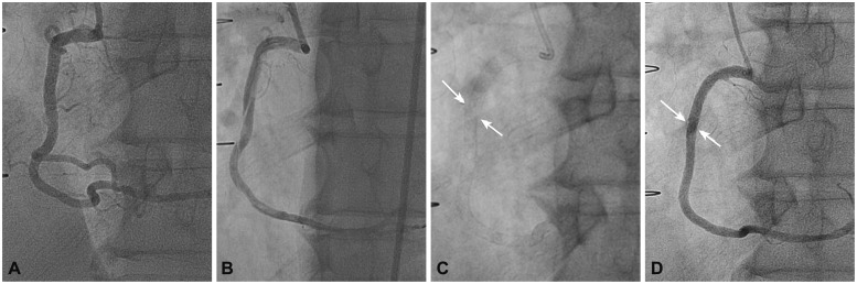
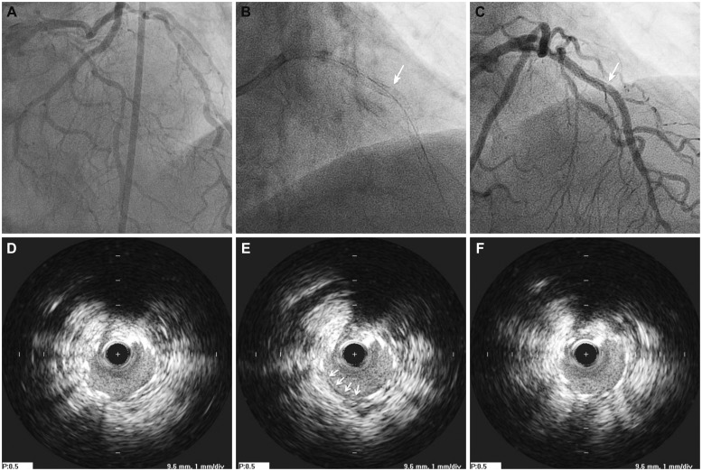
Drug-eluting stent (DES) implantation is currently the standard treatment for various types of coronary artery disease. However, previous reports indicate that stent fractures, which usually occur after a period of time from the initial DES implantation, have increased during the DES era; stent fractures can contribute to unfavorable events such as in-stent restenosis and stent thrombosis. In our present report, we describe two cases of zotarolimus-eluting stent fracture: one that was detected six hours after implementation, and the other case that was detected immediately after deployment. Both anatomical and technical risk factors contributed to these unusual cases of immediate stent fracture.
Keywords: Complications; Drug-eluting stents; Percutaneous coronary intervention.
Conflict of interest statement
The authors have no financial conflicts of interest.
Figures
Similar articles
-
Zotarolimus-eluting stent fracture at initial implantation diagnosed with StentBoost.SAGE Open Med Case Rep. 2016 May 11;4:2050313X16645754. doi: 10.1177/2050313X16645754. eCollection 2016. SAGE Open Med Case Rep. 2016. PMID: 27489714 Free PMC article.
-
Recent progress in percutaneous coronary intervention: evolution of the drug-eluting stents, focus on the XIENCE V drug-eluting stent.Coron Artery Dis. 2010 Jan;21(1):46-56. doi: 10.1097/MCA.0b013e328333f550. Coron Artery Dis. 2010. PMID: 19952925 Review.
-
Differential clinical outcomes after 1 year versus 5 years in a randomised comparison of zotarolimus-eluting and sirolimus-eluting coronary stents (the SORT OUT III study): a multicentre, open-label, randomised superiority trial.Lancet. 2014 Jun 14;383(9934):2047-2056. doi: 10.1016/S0140-6736(14)60405-0. Epub 2014 Mar 14. Lancet. 2014. PMID: 24631162 Clinical Trial.
-
Prospective randomized comparison of clinical and angiographic outcomes between everolimus-eluting vs. zotarolimus-eluting stents for treatment of coronary restenosis in drug-eluting stents: intravascular ultrasound volumetric analysis (RESTENT-ISR trial).Eur Heart J. 2016 Dec 1;37(45):3409-3418. doi: 10.1093/eurheartj/ehw389. Epub 2016 Sep 15. Eur Heart J. 2016. PMID: 27634828
-
Stent thrombosis with drug-eluting stents: is the paradigm shifting?J Am Coll Cardiol. 2013 Nov 19;62(21):1915-1921. doi: 10.1016/j.jacc.2013.08.725. Epub 2013 Sep 11. J Am Coll Cardiol. 2013. PMID: 24036025 Review.
Cited by
-
Zotarolimus-eluting stent fracture at initial implantation diagnosed with StentBoost.SAGE Open Med Case Rep. 2016 May 11;4:2050313X16645754. doi: 10.1177/2050313X16645754. eCollection 2016. SAGE Open Med Case Rep. 2016. PMID: 27489714 Free PMC article.
References
-
- Kuramitsu S, Iwabuchi M, Haraguchi T, et al. Incidence and clinical impact of stent fracture after everolimus-eluting stent implantation. Circ Cardiovasc Interv. 2012;5:663–671. - PubMed
-
- Chakravarty T, White AJ, Buch M, et al. Meta-analysis of incidence, clinical characteristics and implications of stent fracture. Am J Cardiol. 2010;106:1075–1080. - PubMed
-
- Aoki J, Nakazawa G, Tanabe K, et al. Incidence and clinical impact of coronary stent fracture after sirolimus-eluting stent implantation. Catheter Cardiovasc Interv. 2007;69:380–386. - PubMed
-
- Lee MS, Jurewitz D, Aragon J, Forrester J, Makkar RR, Kar S. Stent fracture associated with drug-eluting stents: clinical characteristics and implications. Catheter Cardiovasc Interv. 2007;69:387–394. - PubMed
-
- Kim HS, Kim YH, Lee SW, et al. Incidence and predictors of drug-eluting stent fractures in long coronary disease. Int J Cardiol. 2009;133:354–358. - PubMed
LinkOut - more resources
Full Text Sources
Other Literature Sources
