

Clinico-Pathological Correlation of β-Catenin and Telomere Dysfunction in Head and Neck Squamous Cell Carcinoma Patients
- PMID: 25653721
- PMCID: PMC4314668
- DOI: 10.7150/jca.9558
Clinico-Pathological Correlation of β-Catenin and Telomere Dysfunction in Head and Neck Squamous Cell Carcinoma Patients
Abstract
Background: Tumorigenesis is a complex process of accumulated alteration in function of multiple genes and pathways. Wnt signalling pathway is involved in various differentiation events during embryonic development and is conserved in various species.
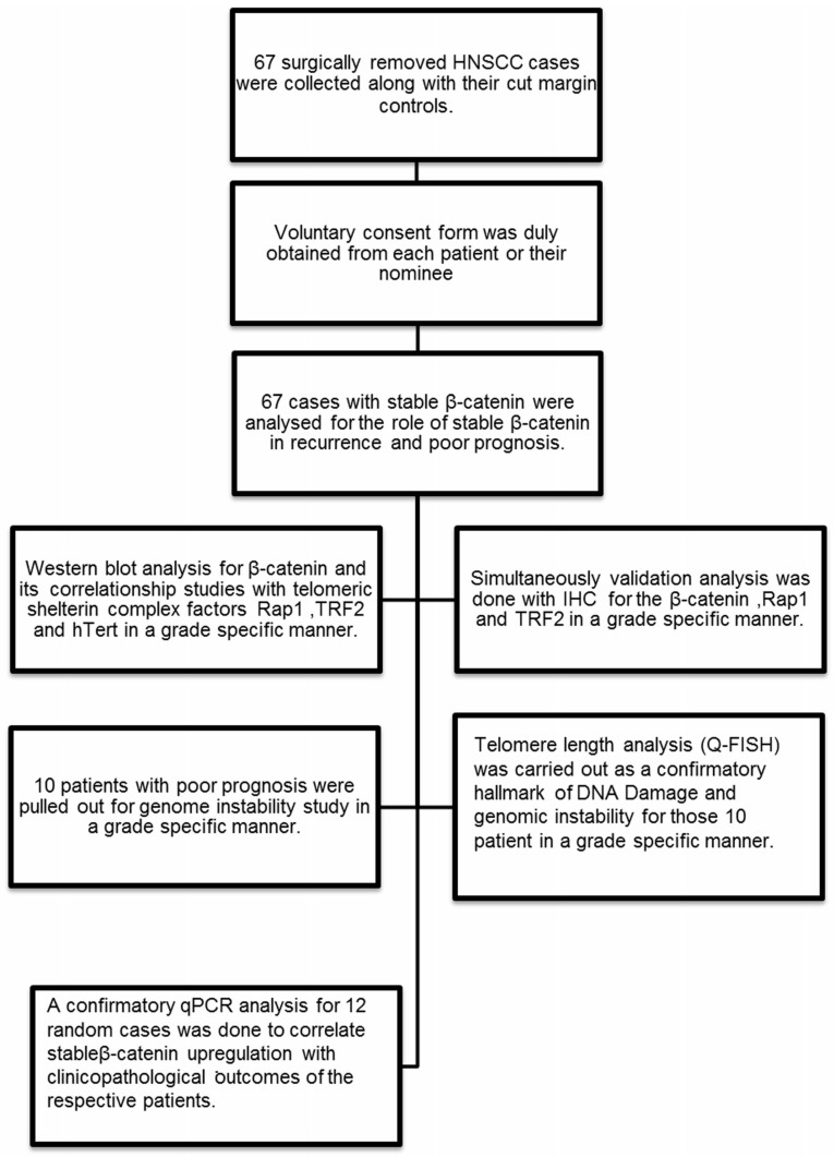
Objective: A multicentre collaborative initiative is undertaken to study the occurrence, prognosis and molecular mechanism of HNSCC (Head and Neck Squamous Cell Carcinoma) which is highly prevalent in eastern parts of India. From a large cohort of HNSCC tissue repository, 67 cases were selected for multi-parametric investigation.
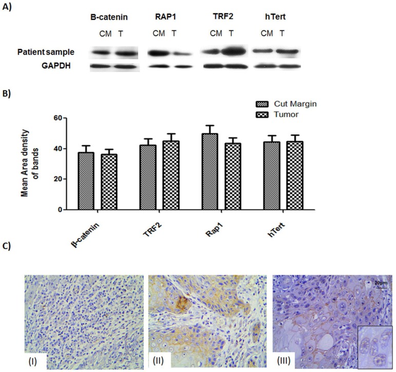
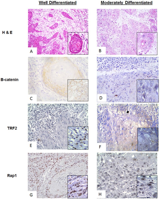
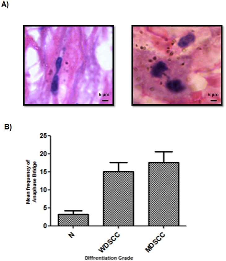
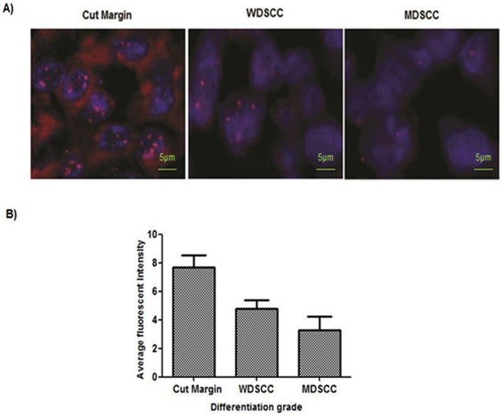
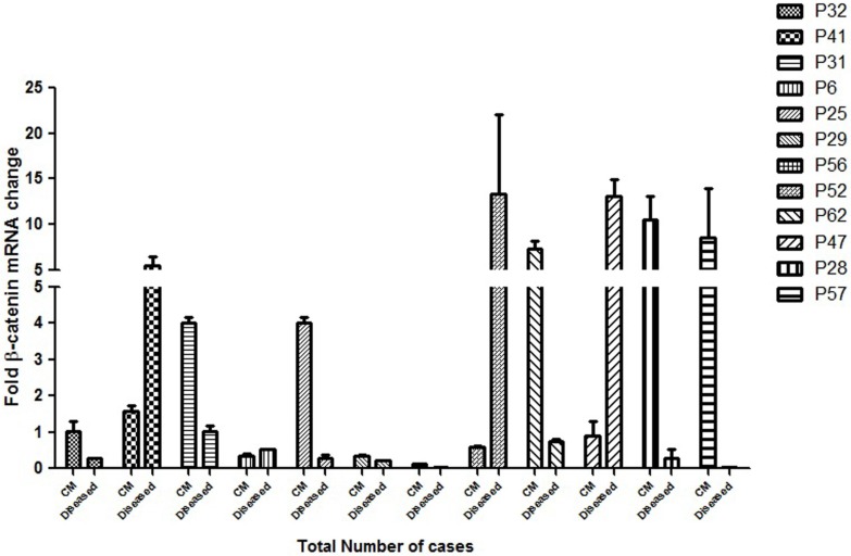
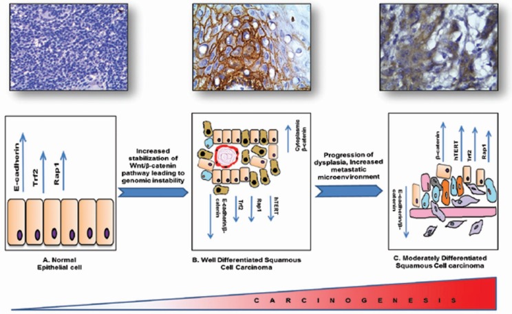
Results: 67 cases showed stable β-catenin expression. We have seen correlation, if any, of the transcription factor - β-catenin, telomere maintenance and shelterin complex proteins - TRF2, Rap1 and hTert with respect to tumor differentiation and telomere dysfunction. Immunohistochemistry of β-catenin protein showed stable and high expression in tumor when compared to stroma. MDSCC (Moderately Differentiated Squamous cell carcinoma) cases expressed nuclear expression of β-catenin in invasive fronts and showed increased genomic instability. Higher frequency of Anaphase bridges was observed ranging from <3% in normal cut margin to 13% in WDSCC (Well differentiated squamous cell carcinoma) and 18% in MDSCC (Moderately differentiated Squamous cell carcinoma). There was significant decrease in telomere length in MDSCC (<4) when compared to the normal cut margin samples (<7). Quantitative Real Time-PCR confirmed a significant correlationship between stable β-catenin expression and poor clinical and pathological outcome.
Conclusion: The Stabilisation and accumulation of β-catenin was significant and correlated well with de-differentiation process as well as prognosis and therapy outcome of the patients in the cohort. Expression status of molecular markers such as β-catenin, hTert, TRF2 and RAP1 correlate significantly with the process of tumorigenesis and prognosis and may play a role in therapeutic management of Head and neck patients.
Keywords: Dedifferentiation; Genomic instability; HNSCC; MDSCC; WDSCC.; Wnt/β-catenin.
Conflict of interest statement
Conflict of Interest: The authors declare no conflict of interest.
Figures
Similar articles
-
Wnt/β-catenin signalling maintains self-renewal and tumourigenicity of head and neck squamous cell carcinoma stem-like cells by activating Oct4.J Pathol. 2014 Sep;234(1):99-107. doi: 10.1002/path.4383. Epub 2014 Jul 23. J Pathol. 2014. PMID: 24871033
-
Role of β-catenin in cisplatin resistance, relapse and prognosis of head and neck squamous cell carcinoma.Cell Oncol (Dordr). 2018 Apr;41(2):185-200. doi: 10.1007/s13402-017-0365-1. Epub 2017 Dec 14. Cell Oncol (Dordr). 2018. PMID: 29243047
-
Rap1 stabilizes beta-catenin and enhances beta-catenin-dependent transcription and invasion in squamous cell carcinoma of the head and neck.Clin Cancer Res. 2010 Jan 1;16(1):65-76. doi: 10.1158/1078-0432.CCR-09-1122. Epub 2009 Dec 22. Clin Cancer Res. 2010. PMID: 20028760 Free PMC article.
-
Telomeres and telomerase in head and neck squamous cell carcinoma: from pathogenesis to clinical implications.Cancer Metastasis Rev. 2016 Sep;35(3):457-74. doi: 10.1007/s10555-016-9633-1. Cancer Metastasis Rev. 2016. PMID: 27501725 Free PMC article. Review.
-
Calprotectin and the Initiation and Progression of Head and Neck Cancer.J Dent Res. 2018 Jun;97(6):674-682. doi: 10.1177/0022034518756330. Epub 2018 Feb 14. J Dent Res. 2018. PMID: 29443623 Free PMC article. Review.
Cited by
-
Prolonged inflammatory microenvironment is crucial for pro-neoplastic growth and genome instability: a detailed review.Inflamm Res. 2017 Feb;66(2):119-128. doi: 10.1007/s00011-016-0985-3. Epub 2016 Sep 21. Inflamm Res. 2017. PMID: 27653961 Review.
-
Role of Telomeric TRF2 in Orosphere Formation and CSC Phenotype Maintenance Through Efficient DNA Repair Pathway and its Correlation with Recurrence in OSCC.Stem Cell Rev Rep. 2018 Dec;14(6):871-887. doi: 10.1007/s12015-018-9823-z. Stem Cell Rev Rep. 2018. PMID: 29872959
-
p38 MAPK pathway and its interaction with TRF2 in cisplatin induced chemotherapeutic response in head and neck cancer.Oncogenesis. 2018 Jul 9;7(7):53. doi: 10.1038/s41389-018-0062-6. Oncogenesis. 2018. PMID: 29983416 Free PMC article.
-
Differences in Extracellular Vesicle Protein Cargo Are Dependent on Head and Neck Squamous Cell Carcinoma Cell of Origin and Human Papillomavirus Status.Cancers (Basel). 2021 Jul 23;13(15):3714. doi: 10.3390/cancers13153714. Cancers (Basel). 2021. PMID: 34359613 Free PMC article.
-
Emerging Insights into Wnt/β-catenin Signaling in Head and Neck Cancer.J Dent Res. 2018 Jun;97(6):665-673. doi: 10.1177/0022034518771923. J Dent Res. 2018. PMID: 29771197 Free PMC article. Review.
References
-
- Saranath D. Cancer: a global view. Contemporary Issues in Oral Cancer. Oxford University Press. 2000. pp. 1–29.
-
- Nair U, Bartsch H, Nair J. Alert for an epidemic of oral cancer due to use of the betel quid substitute's gutkha and pan masala: a review of agents and causative mechanisms. Mutagenesis. 2004;19:251–262. - PubMed
LinkOut - more resources
Full Text Sources
Other Literature Sources
Research Materials
Miscellaneous
