

Effects of BIO on proliferation and chondrogenic differentiation of mouse marrow-derived mesenchymal stem cells
- PMID: 25653775
- PMCID: PMC4313005
Effects of BIO on proliferation and chondrogenic differentiation of mouse marrow-derived mesenchymal stem cells
Abstract
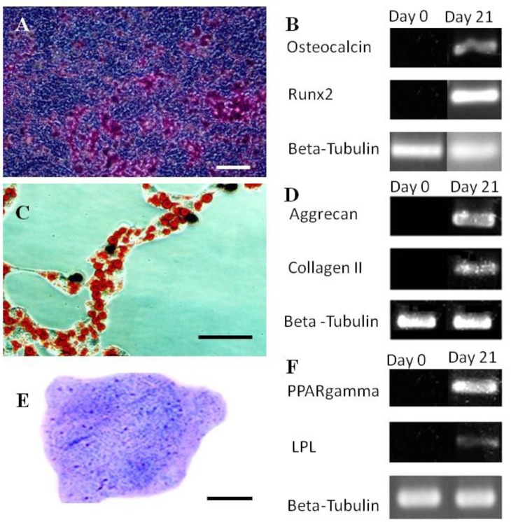
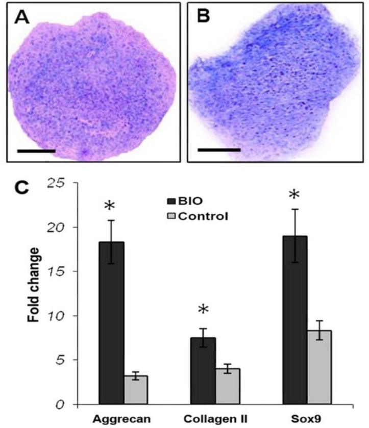
In vitro expansion of mesenchymal stem cell (MSCs) into large number is necessary for their application in cell-based treatment of articular cartilage defects. On the other hand, some studies have indicated that BIO (6-Bromoindirubin-3-Oxime) possesses mitogenic effects on cell culture. The objective of the present study was to examine the effect of BIO on in vitro expansion and chondrogenic differentiation of mouse marrow-derived MSCs. The culture was established using bone marrow tissue obtained from 10 NMRI mice. MSC nature of the isolated cells was verified according to the minimal criteria proposed for MSC. Passaged-3 cells were seeded in 24-well culture plates and treated by 0.05, 0.01, 0.1, 1.0 and 1.5 µM BIO for seven days. The culture without BIO was taken as the control. At the end of cultivation period, the cultures were examined for viable cell number which was then used to calculate population doubling time (PDT). The BIO with higher proliferation-promoting effect was investigated for its chondrogenic effect on MSC culture. There was significantly more viable cells at the cultures treated by 0.1 µM BIO. At this culture the cells tended to double their population in rapid rate (each 43.07 hr) than the cells treated with the other BIO concentrations (p < 0.05). Interestingly treatment of MSC chondrogenic culture with 0.1 µM BIO led to the up-regulation of cartilage specific genes including aggrecan, collagen II and Sox9. In conclusion BIO at 0.1 µM could enhance mouse MSC in vitro proliferation as well as their chondrogenic differentiation. These findings would be of great importance for the field of regenerative medicine.
Keywords: BIO; Cartilage differentiation; Mesenchymal stem cells; Mouse; Proliferation.
Figures


Similar articles
-
Small Molecule-BIO Accelerates and Enhances Marrow-Derived Mesenchymal Stem Cell in Vitro Chondrogenesis.Iran J Med Sci. 2014 Mar;39(2):107-16. Iran J Med Sci. 2014. PMID: 24644379 Free PMC article.
-
The Effects of the WNT-Signaling Modulators BIO and PKF118-310 on the Chondrogenic Differentiation of Human Mesenchymal Stem Cells.Int J Mol Sci. 2018 Feb 13;19(2):561. doi: 10.3390/ijms19020561. Int J Mol Sci. 2018. PMID: 29438298 Free PMC article.
-
Chondrogenic differentiation of bovine bone marrow mesenchymal stem cells (MSCs) in different hydrogels: influence of collagen type II extracellular matrix on MSC chondrogenesis.Biotechnol Bioeng. 2006 Apr 20;93(6):1152-63. doi: 10.1002/bit.20828. Biotechnol Bioeng. 2006. PMID: 16470881
-
Coculture of equine mesenchymal stem cells and mature equine articular chondrocytes results in improved chondrogenic differentiation of the stem cells.Jpn J Vet Res. 2010 May;58(1):5-15. Jpn J Vet Res. 2010. PMID: 20645581
-
Repair of injured articular and growth plate cartilage using mesenchymal stem cells and chondrogenic gene therapy.Curr Stem Cell Res Ther. 2006 May;1(2):213-29. doi: 10.2174/157488806776956904. Curr Stem Cell Res Ther. 2006. PMID: 18220868 Review.
References
-
- Pittenger MF, Mackay AM, Beck SC, et al. Multilineage potential of adult human mesenchymal stem cells. Science. 1999;248(5411):143–147. - PubMed
-
- Eslaminejad MB, Nikmahzar A, Taghiyar L, et al. Murine mesenchymal stem cells isolated by low density primary culture system. Dev Growth Differ. 2006;48(6):361–370. - PubMed
-
- Friedenstein AJ, Chailakhjan RK, Lalykina KS. The development of fibroblast colonies in monolayer cultures of guinea pig bone marrow and spleen cells. Cell Tissue Kinet. 1970;3:393–403. - PubMed
-
- Friedenstein AJ, Gorskaja JF, Kulagina NN. Fibroblast precursors in normal and irradiated mouse hematopoietic organs. Exp Hematol. 1976;3(4):267–274. - PubMed
-
- Owen M. Histogenesis of bone cells. Calcif Tissue Res. 1978;25(3):205–207. - PubMed
LinkOut - more resources
Full Text Sources
Research Materials