Review
. 2015:2015:606479.
doi: 10.1155/2015/606479. Epub 2015 Jan 11.

Sweet taste receptor signaling network: possible implication for cognitive functioning

Affiliations
Review

Sweet taste receptor signaling network: possible implication for cognitive functioning

Menizibeya O Welcome et al. Neurol Res Int. 2015.

Abstract

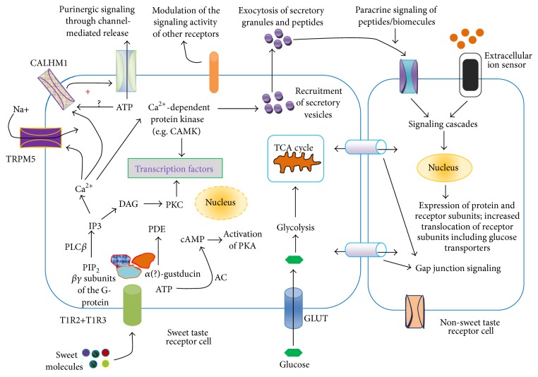
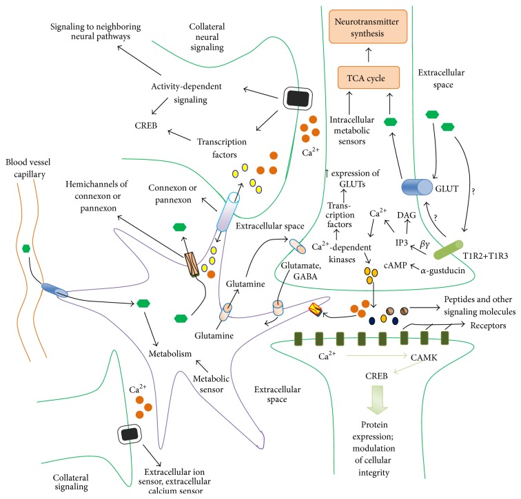
Sweet taste receptors are transmembrane protein network specialized in the transmission of information from special "sweet" molecules into the intracellular domain. These receptors can sense the taste of a range of molecules and transmit the information downstream to several acceptors, modulate cell specific functions and metabolism, and mediate cell-to-cell coupling through paracrine mechanism. Recent reports indicate that sweet taste receptors are widely distributed in the body and serves specific function relative to their localization. Due to their pleiotropic signaling properties and multisubstrate ligand affinity, sweet taste receptors are able to cooperatively bind multiple substances and mediate signaling by other receptors. Based on increasing evidence about the role of these receptors in the initiation and control of absorption and metabolism, and the pivotal role of metabolic (glucose) regulation in the central nervous system functioning, we propose a possible implication of sweet taste receptor signaling in modulating cognitive functioning.


Figures

Figure 1
Figure 1
A general model of sweet taste signaling network. Sweet taste receptors possess multiple binding sites and mode of interaction for sweet molecules and they belong to class C of heterotrimeric guanine nucleotide-binding protein, G-protein [–145]. Sweet molecules activate the G-protein by downstream signaling leading to the dissociation of the α-subunit gustducin from the βγ subunits [146, 147]. Dissociated βγ subunits of the G-protein activate phospholipase Cβ (PLCβ), leading to the formation of 1,4,5-inositol trisphosphate (IP3). IP3 is responsible for the release of Ca2+ from intracellular stores through its binding to IP3-receptor in these stores. Increase in intracellular Ca2+ activates calcium dependent kinase, monovalent selective cation channel, TRPM5 (transient receptor potential cation channel, subfamily M, member 5) [15, 44, 146], and other receptors [44, 148]. To establish the role of TRPM5 or PLCβ (type 2), Zhang et al. [4] showed that knockout of the receptor or the enzyme abolishes the sensation of taste in cells. TRPM5 may also play a role in capacitance mediated calcium entry into taste cells [147]. Modulation of purinergic signaling by taste receptor also plays useful role in taste sensation. For the initiation of purinergic release, it was recently demonstrated by Taruno et al. [148] that the voltage-gated ion channel, calcium homeostasis modulator 1 (CALHM1), is indispensable for taste-stimuli-evoked ATP release from sweet, bitter, and umami taste cells. Importantly, CALHM1 is expressed not only in sweet but also in bitter and umami taste sensing type 2 cells. Taruno et al. [148] proposed that CALHM1 is a voltage-gated ATP-release channel. Dissociated α subunit referred to as Gα-gustducin activates a phosphodiesterase (PDE) thereby decreasing intracellular cAMP levels [146, 149]. Gα-gustducin is also reported to activate adenylate cyclase (AC) to increase cAMP level [150]. According to earlier report, Clapp et al. [151] demonstrated that, compared to wild type mice, knockout of α-gustducin in the taste buds of mice resulted in high basal (unstimulated) cAMP level. The results of these authors [151] indicated that α-gustducin is necessary to maintain low level of cAMP level. Low level of cAMP is necessary to maintain the adequate signaling of Ca2+ by disinhibition of cyclic nucleotide-inhibited channels to elevate intracellular Ca2+ [38]. Changes in cAMP levels also affect other ion channels, including K+ channels. The events resulting in activation/modulation of ion channels lead to membrane depolarization and formation of action potentials. Potential-dependent release of mediators (ATP, serotonin, etc.) and peptides and calcium dependent release of peptides/biomolecules are some of the results of sweet taste receptor signaling [152]. A hallmark of sweet taste receptor signaling is the activation of transcription factors and gene expression, which might be dependent on calcium and activity dependent activation calcium dependent kinases, including the calmodulin-dependent protein kinase (CAMK). Activation of protein kinases may be achieved through other signaling pathways. It appears that sweet taste receptor signaling involves multiple activating substrates and different types and subtypes of both α-gustducin and βγ subunits of the G-protein. Although, different subtypes of sweet taste G-protein receptor subunits have been known for over a decade, their specific roles in sensing taste are not exactly clear [38, 149, 153]. For instance, Huangu et al. [149] reported the presence of β1 and γ13. The sweet taste receptor is also known to have β3 subtype subunit. For α-gustducin, Gα i-2, Gα i-3, 14, 15, Gα q, Gα s, α-transducin have been identified [38].
Figure 2
Figure 2
Sweet taste signaling network of the neuroastroglial system. The brain is a complex network of cells, largely populated by neurons and astrocytes. There are ~100 billion neurons, with glial cells outnumbering neurons by about 10-fold. Astrocytes form the largest population of glial cells. The metabolic role of astrocytes in brain has been reviewed in our previous work [60]. Mechanisms of how glucose enters into the astrocytes and neurons are well documented [61]. From the scheme (Figure 2), the presynaptic neuron senses glucose mediated by the T1R2+T1R3 and GLUT2. While the mechanisms, by which the neuron senses glucose through GLUT2, have increasingly been defined, the glucosensing mechanisms of the sweet taste receptor are yet to be understood. It is quite possible that sweet taste receptor can modulate the plasma membrane GLUT2 glucosensor. Functioning cooperatively with GLUT2 to sense the metabolic rate of the intracellular milieu is the G-protein coupled receptor, the inwardly rectifying ATP-dependent potassium channel (KATP channel) [62, 63]. Transport activity of GLUT2 may be affected through multiple signaling pathways, such as those involving the regulation of GLUT2 and KATP channel activity. While in Ren et al. [24] study, the signaling activity of GLUT2 was not assessed, their results showed that the inhibition of sweet taste receptor resulted in increase in taste receptor gene expression, suggesting that sweet taste receptors persistently code information about the extracellular glucose level to intracellular milieu, and this might, probably, involve intracellular metabolic sensors, mediating neural activity, gene expression, and membrane receptor trafficking. Although, the mechanisms of the T1R2+T1R3/GLUT2-cooperativity/associativity (or intracellular metabolic sensors) interaction are not known, it can be proposed that T1R2+T1R3 could modulate GLUT2 transport activity through mechanisms as yet unknown. Mechanism of downstream signaling of the neuronal T1R2+T1R3 receptor is similar to that in other cells (Figure 1). The downstream signaling of these receptors can result in changes in extracellular calcium concentration as well as changes in peptide secretions. These biomolecules are sensed by their corresponding receptors in the adjacent neurons/astrocytes, which couple the received information into intracellular signal and cellular activity. The waves of calcium ions, peptide-dependent signaling, can affect collateral neurons and astrocytes by activity dependent signaling and changes in ion waves and regulate gene expression and protein synthesis. Several transcription factors and memory relation genes are activated/deactivated. Intercellular signaling through connexons and pannexons in these cells can modulate their activity.

Similar articles

Cited by

References

    1. Nelson G., Hoon M. A., Chandrashekar J., Zhang Y., Ryba N. J. P., Zuker C. S. Mammalian sweet taste receptors. Cell. 2001;106(3):381–390. doi: 10.1016/S0092-8674(01)00451-2. - DOI - PubMed
    1. Adler E., Hoon M. A., Mueller K. L., Chandrashekar J., Ryba N. J. P., Zuker C. S. A novel family of mammalian taste receptors. Cell. 2000;100(6):693–702. doi: 10.1016/S0092-8674(00)80705-9. - DOI - PubMed
    1. Sainz E., Cavenagh M. M., LopezJimenez N. D., et al. The G-protein coupling properties of the human sweet and amino acid taste receptors. Developmental Neurobiology. 2007;67(7):948–959. doi: 10.1002/dneu.20403. - DOI - PubMed
    1. Zhang Y., Hoon M. A., Chandrashekar J., et al. Coding of sweet, bitter, and umami tastes: different receptor cells sharing similar signaling pathways. Cell. 2003;112(3):293–301. doi: 10.1016/s0092-8674(03)00071-0. - DOI - PubMed
    1. Blad C. C., Tang C., Offermanns S. G protein-coupled receptors for energy metabolites as new therapeutic targets. Nature Reviews Drug Discovery. 2012;11(8):603–619. doi: 10.1038/nrd3777. - DOI - PubMed

LinkOut - more resources