Review
. 2015 Jan 21:2:148.
doi: 10.3389/fped.2014.00148. eCollection 2014.

The lectin pathway of complement and rheumatic heart disease

Affiliations
Review

The lectin pathway of complement and rheumatic heart disease

Marcia Holsbach Beltrame et al. Front Pediatr. .

Abstract

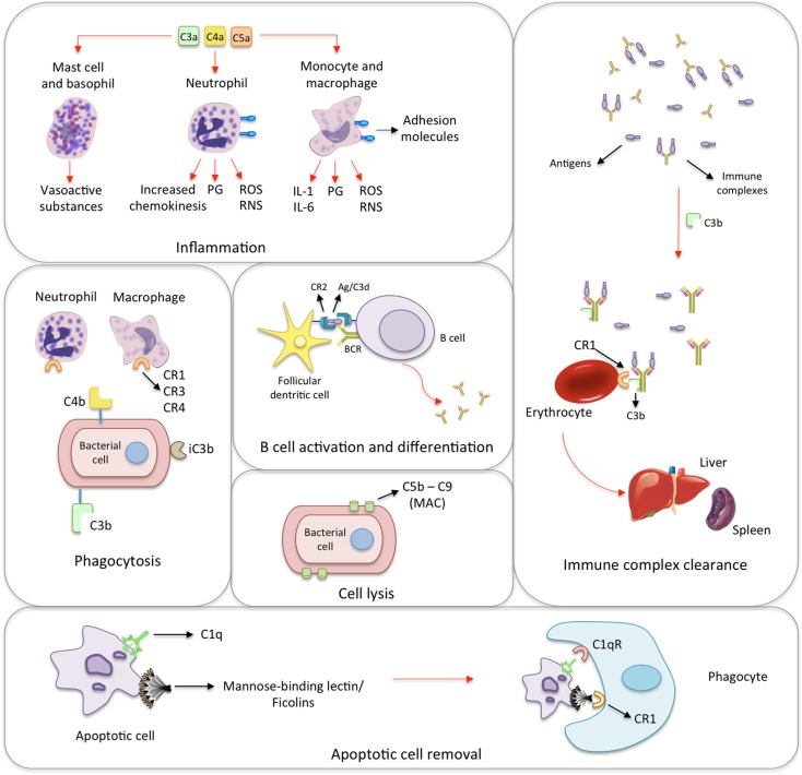
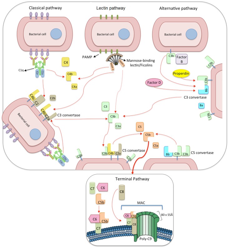
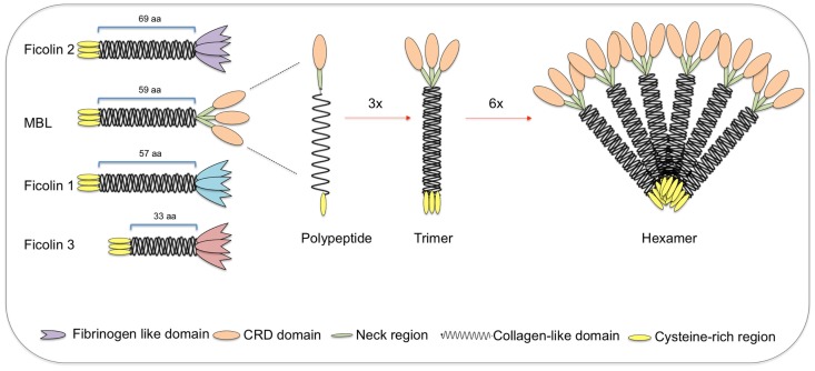
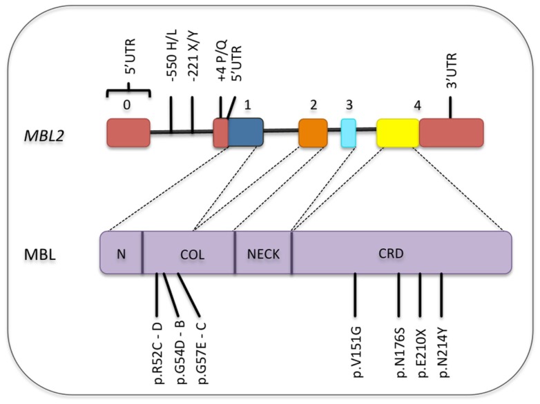
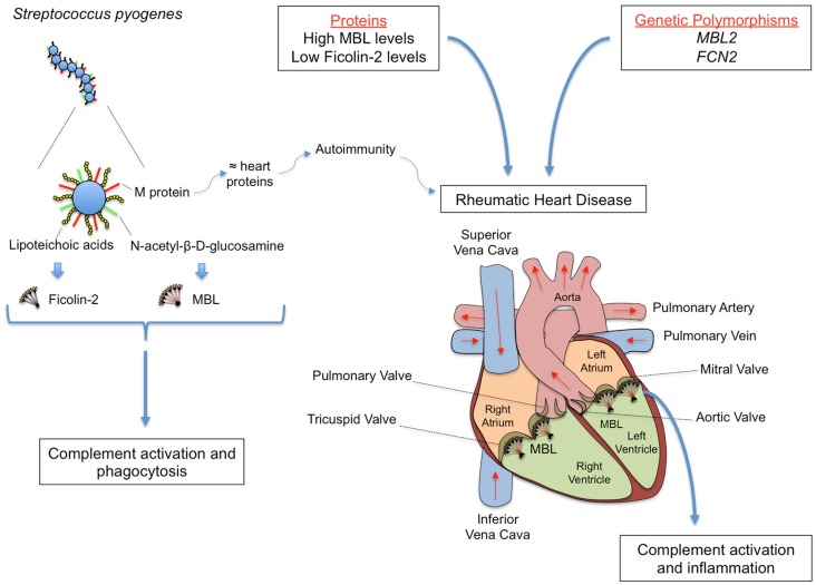
The innate immune system is the first line of host defense against infection and is comprised of humoral and cellular mechanisms that recognize potential pathogens within minutes or hours of entry. The effector components of innate immunity include epithelial barriers, phagocytes, and natural killer cells, as well as cytokines and the complement system. Complement plays an important role in the immediate response against microorganisms, including Streptococcus sp. The lectin pathway is one of three pathways by which the complement system can be activated. This pathway is initiated by the binding of mannose-binding lectin (MBL), collectin 11 (CL-K1), and ficolins (Ficolin-1, Ficolin-2, and Ficolin-3) to microbial surface oligosaccharides and acetylated residues, respectively. Upon binding to target molecules, MBL, CL-K1, and ficolins form complexes with MBL-associated serine proteases 1 and 2 (MASP-1 and MASP-2), which cleave C4 and C2 forming the C3 convertase (C4b2a). Subsequent activation of complement cascade leads to opsonization, phagocytosis, and lysis of target microorganisms through the formation of the membrane-attack complex. In addition, activation of complement may induce several inflammatory effects, such as expression of adhesion molecules, chemotaxis and activation of leukocytes, release of reactive oxygen species, and secretion of cytokines and chemokines. In this chapter, we review the general aspects of the structure, function, and genetic polymorphism of lectin-pathway components and discuss most recent understanding on the role of the lectin pathway in the predisposition and clinical progression of Rheumatic Fever.

Keywords: MBL; complement system; ficolins; gene polymorphisms; lectin pathway.


Figures

Figure 1
Figure 1
Biological functions of the complement system. Inflammation: the activation of the complement system generates many anaphylatoxins, among which C3a, C4a, and C5a. The binding of C3a, C4a, and C5a to receptors on mast cells and basophils leads to the release of histamine and other vasoactive mediators. In response to the activation by anaphylatoxins, neutrophils release prostaglandins (PG), reactive oxygen and nitrogen species (ROS and RNS, respectively) as well as increase the expression of adhesion molecules, and chemokinesis. Monocytes and macrophages show similar response and secrete interleukins 1 and 6 (IL-1 and IL-6). Phagocytosis: iC3b, C4b, and mainly C3b coat microorganisms and immune complexes, having opsonizing activity. Neutrophils and macrophages express complement receptors (CR1, CR2, and CR4), which bind C3b, C4b, and iC3b. This promotes the adherence of the microorganism to phagocytic host cells leading to phagocytosis. B cell activation and differentiation: the recognition of C3-tagged antigen plays an important role in B cell activation and differentiation. Cross-linking between complement receptor 2 (CR2) and B cell receptor (BCR) through C3d–antigen complexes lowers the threshold of B cell activation leading to migration, T cell/B cell interaction and antibody class-switch. Cell lysis: specific antibodies, MBL/ficolins, and spontaneous hydrolysis of C3 activate the complement on the surface of infectious microorganisms and lead to the formation of membrane-attack complexes (MAC), which cause their lysis. Immune complex clearance: immune complexes activate the complement system. The generated C3b binds to the complexes and to CR1 present on the surface of erythrocytes. During erythrocyte traffic through sinusoids in liver and spleen, resident phagocytes remove bound immune complexes leading to their clearance. Apoptotic cell removal: Mannose-binding lectin, ficolins and C1q bind debris of apoptotic cells, which are subsequently removed through binding to the C1qR and CR1 receptors on phagocytic cells.
Figure 2
Figure 2
The three pathways of complement activation: classical, lectin, and alternative pathways. The classical pathway is initiated via binding of C1 complex (which consists of C1q, C1r, and C1s molecules) through its recognition molecule C1q to antibody complexes on the surface of pathogens. Subsequently, C1s cleaves C4, which binds covalently to the pathogen surface, and then cleaves C2, leading to the formation of C4b2a complex, the C3 convertase of the classical pathway. Activation of the lectin pathway occurs through the binding of the complex of mannose-binding lectin (MBL), CL-K1 or ficolins, and MBL-associated serine proteases 1 and 2 (MASP-1 and MASP-2, respectively) to various carbohydrates or acetylated residues on the surface of pathogens (PAMP, pathogen-associated molecular pattern). Like C1s, MASP-2 leads to the formation of the C3 convertase, C4b2a, but its activation is dependent on MASP-1. MASP-1 also cleaves C2 and C3. Activation of the alternative pathway depends on spontaneous low-grade hydrolysis of C3 in plasma leading to the formation of C3b. This C3b binds factor B (homologous to C2) to form a C3bB complex. The cleavage of factor B by factor D form the alternative pathway C3 convertase, C3bBb. Properdin stabilizes this complex. The C3 convertases cleave C3 to C3b, which binds covalently next to the site of complement activation (opsonization). This amplifies the cascade and mediates phagocytosis, as well as adaptative immune responses. The addition of further C3b molecules to the C3 convertase forms C5 convertases (C3bBbC3b for the alternative pathway or C4bC2aC3b for both classical and lectin pathways), initiating the assembly of the membrane-attack complex (MAC) by cleavage of C5 to C5a and C5b. Whereas C5a functions as a potent anaphylatoxin, C5b forms a complex with C6 and C7, which is inserted in the cell membrane. Thereafter, C8 and 10–18 C9 molecules (80 × 55 Å each) bind to this complex, resulting in a fully functional MAC (C5b-9). The three pathways converge to this common terminal pathway, culminating with cell lysis and death.
Figure 3
Figure 3
Structural subunits of mannan-binding lectin (MBL) and ficolins. Both MBL and ficolins contain a short N-terminal cysteine-rich region followed by a collagen-like sequence [length given in number of amino acids (aa)]. The C-terminal region is a carbohydrate-recognition binding domain for MBL (shown as oval forms) and a fibrinogen-like domain for ficolins (shown as tulip forms). The polypeptides interact through its collagen-like region forming triple helices (trimers), which further associate into higher oligomeric arrangements (tetramers to hexamers). Despite the very different structures, ficolin polypeptides and trimers interact in the same way as MBL, forming high oligomeric forms (tetramers). MBL-associated serine proteases interact with the collagen-like region, thereby activating the lectin pathway of complement.
Figure 4
Figure 4
Common polymorphisms in the MBL2 gene and its corresponding locations in the MBL protein. Only the functional polymorphisms in the promoter and non-synonymous mutations are shown [SNP database and Boldt et al. (161)]. Exons are numbered. Exons, introns, and protein domains are not in scale. N, N-terminal region; COL, collagen-like region; CRD, carbohydrate-recognition domain.
Figure 5
Figure 5
Rheumatic fever (RF) and its most severe sequel chronic rheumatic heart disease (RHD) are chronic inflammations that follow oropharynx infection by Streptococcus pyogenes, whose cell wall presents several PAMPs, including M protein, lipoteichoic acid, and N-acetil-β-d-glucosamine. M protein shares structural homology with heart proteins such as myosin and tropomyosin, leading to the formation of cross-reactive auto-antibodies. MBL and Ficolin-2 bind N-acetyl-β-d-glucosamine and lipoteichoic acid, respectively, inducing complement activation and phagocytosis. Although conferring protection against the initial infection, MBL may deposit on the altered valves, eliciting inflammation, and complement tissue damage in the chronic stage of the disease. The functional importance of the proteins may vary during infection and disease establishment, with MBL2 and FCN2 polymorphisms leading to high MBL and low Ficolin-2 levels, respectively, being associated with increased susceptibility to RHD.

Similar articles

Cited by

References

    1. Dunkelberger JR, Song W-C. Complement and its role in innate and adaptive immune responses. Cell Res (2010) 20:34–50.10.1038/cr.2009.139 - DOI - PubMed
    1. Walport MJ. Complement. First of two parts. N Engl J Med (2001) 344:1058–6610.1056/NEJM200104053441406 - DOI - PubMed
    1. Degn SE, Thiel S. Humoral pattern recognition and the complement system. Scand J Immunol (2013) 78:181–93.10.1111/sji.12070 - DOI - PubMed
    1. Ricklin D, Hajishengallis G, Yang K, Lambris JD. Complement: a key system for immune surveillance and homeostasis. Nat Immunol (2010) 11:785–97.10.1038/ni.1923 - DOI - PMC - PubMed
    1. Blom AM, Villoutreix BO, Dahlbäck B. Complement inhibitor C4b-binding protein-friend or foe in the innate immune system? Mol Immunol (2004) 40:1333–46.10.1016/j.molimm.2003.12.002 - DOI - PubMed