. 2015 Mar 3;87(5):2535-41.
doi: 10.1021/ac504685y. Epub 2015 Feb 9.

Quantitative chemoproteomics for site-specific analysis of protein alkylation by 4-hydroxy-2-nonenal in cells

Affiliations

Quantitative chemoproteomics for site-specific analysis of protein alkylation by 4-hydroxy-2-nonenal in cells

Jing Yang et al. Anal Chem. .

Abstract

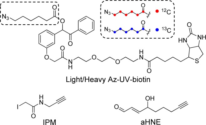
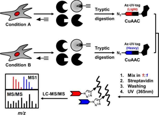
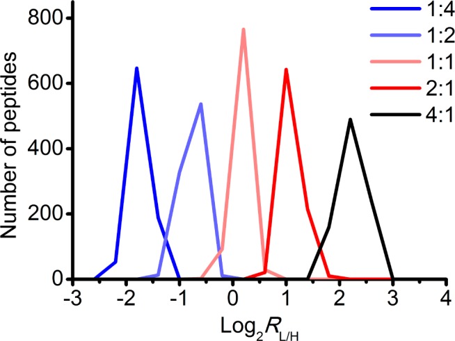
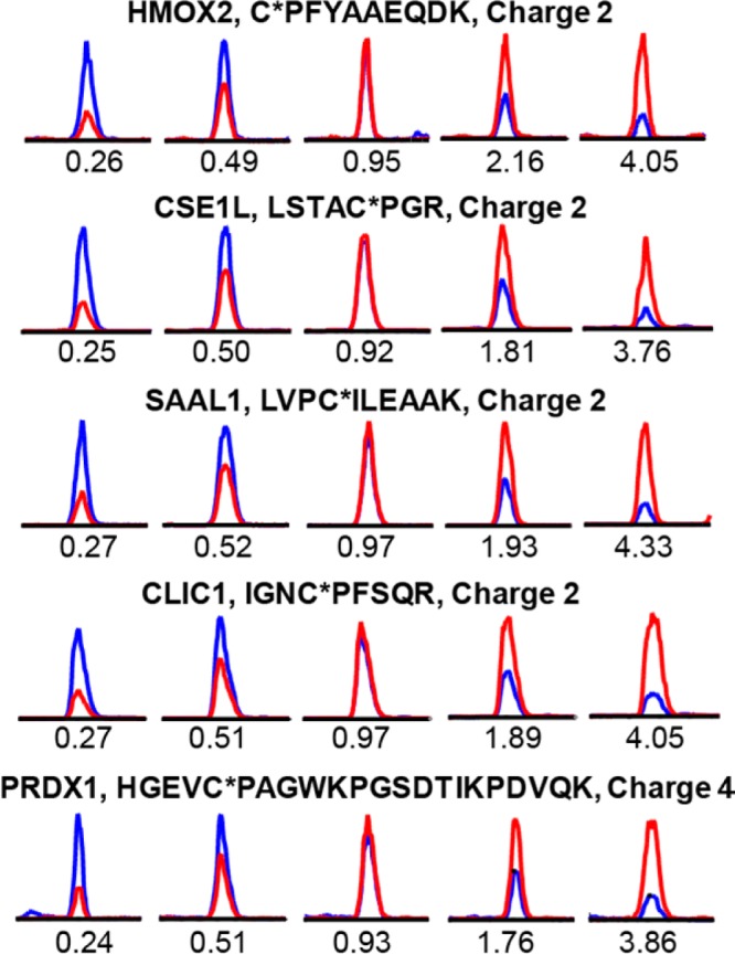
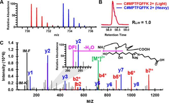
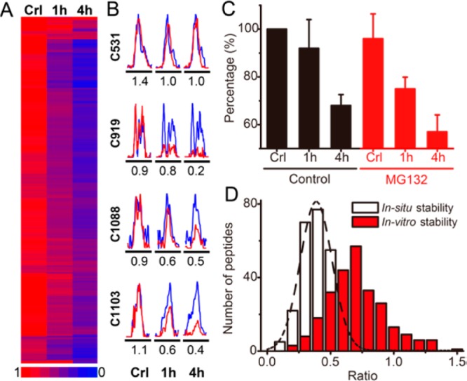
Protein alkylation by 4-hydroxy-2-nonenal (HNE), an endogenous lipid derived electrophile, contributes to stress signaling and cellular toxicity. Although previous work has identified protein targets for HNE alkylation, the sequence specificity of alkylation and dynamics in a cellular context remain largely unexplored. We developed a new quantitative chemoproteomic platform, which uses isotopically tagged, photocleavable azido-biotin reagents to selectively capture and quantify the cellular targets labeled by the alkynyl analogue of HNE (aHNE). Our analyses site-specifically identified and quantified 398 aHNE protein alkylation events (386 cysteine sites and 12 histidine sites) in intact cells. This data set expands by at least an order of magnitude the number of such modification sites previously reported. Although adducts formed by Michael addition are thought to be largely irreversible, we found that most aHNE modifications are lost rapidly in situ. Moreover, aHNE adduct turnover occurs only in intact cells and loss rates are site-selective. This quantitative chemoproteomics platform provides a versatile general approach to map bioorthogonal-chemically engineered post-translational modifications and their cellular dynamics in a site-specific and unbiased manner.


Figures

Scheme 1
Scheme 1. Chemical Structures of Light (Red) and Heavy (Blue) Azido-UV–Biotin Reagents and Alkynyl Electrophile Probes Used in This Study
Figure 1
Figure 1
Schematic representation of a site-centric quantitative chemoproteomic workflow.
Figure 2
Figure 2
Validation of the accuracy of quantitative chemoproteomic analysis. RKO proteomes were labeled with the alkyne tagged cysteine alkylating reagent, IPM (Scheme 1), and digested into tryptic peptides. Aliquots of peptide mixtures were conjugated with light or heavy isotopic tagged Az-UV-biotin reagents and mixed in predefined ratios (RL/H = 1:4, 1:2 1:1, 2:1, 4:1). After affinity capture and photorelease, the alkylated peptides were analyzed by LC–MS/MS, and the light/heavy ratios were calculated for IPM-modified cysteine containing peptides. The distributions of these ratios demonstrate the accuracy of this quantitative chemoproteomic workflow. Data are displayed using a log 2 scale on the x axis.
Figure 3
Figure 3
Representative extracted ion chromatograms (XIC) for the IPM-labeled peptides from five proteins at predefined ratios (L/H = 1:4, 1:2 1:1, 2:1, 4:1, from left to right). The profiles for light- and heavy-labeled peptides are shown in red and blue, respectively. The peptide sequence, modified sites (with asterisk), and charge status are shown above the individual chromatograms. The measured light/heavy ratios (RL/H) are displayed below each individual chromatogram.
Figure 4
Figure 4
Identification of Cys-73 of thioredoxin 1 as an alkylation target site by aHNE in RKO cells. (A) MS1 spectrum of an aHNE-triazol-hexanoic acid modified peptide from thioredoxin 1. Doubly charged monoisotopic precursors of light and heavy labeled the peptide are observed at m/z 730.3588 (red) and 733.3686 (blue), respectively, with mass errors less than 1.0 ppm. (B) XIC are shown for changes in the same aHNE-modified peptides from thioredoxin with the profiles for light- and heavy-labeled peptides in red and blue, respectively. (C) Characteristic fragmentation of the light-labeled modified peptide and its HCD MS/MS spectrum. A zoom window displays the diagnostic fragment ion (DFI) peak (m/z 292.2). The asterisks on the annotated ions indicate water losses from the corresponding b- and y-ion fragments.
Figure 5
Figure 5
Dynamics of protein S-alkylation by aHNE in RKO cells. (A) Heatmap of ratios of changes of all detected cysteine S-alkylation events shows that most adducts turn over rapidly in a time-dependent manner in cells. Lower the measured ratio (L/H) indicates more rapid turnover. (B) XIC are shown for changes in S-alkylated peptides from FAM120A protein in RKO cells, with the profiles for light- and heavy- labeled peptides in red and blue, respectively. The mean measured ratios were calculated from three biological replicate experiments and are displayed below the individual chromatograms, respectively. (C) Turnover of alkylation is not affected by proteasome inhibition. RKO cells were pretreated with (red) or without MG132 (black), followed by aHNE treatment with or without 1 and 4 h recovery periods. Proteins alkylated by aHNE were labeled with azido-biotin and detected by Western blotting with fluorescein-conjugated streptavidin. Data were presented as mean values ± SD, n = 3 biological replicates per group. A representative Western blot is shown in Figure S10 in the Supporting Information. (D) Distributions of the measured ratios of dynamic aHNE-cysteine adduction in vitro (red) and in situ (white).

References

    1. Rudolph T. K.; Freeman B. A. Sci. Signaling 2009, 2, re7. - PMC - PubMed
    1. Dalleau S.; Baradat M.; Gueraud F.; Huc L. Cell Death Differ. 2013, 20, 1615–1630. - PMC - PubMed
    1. Barrera G.; Pizzimenti S.; Ciamporcero E. S.; Daga M.; Ullio C.; Arcaro A.; Cetrangolo G. P.; Ferretti C.; Dianzani C.; Lepore A.; Gentile F. Antioxid. Redox Signaling 2014, 10.1089/ars.2014.6166. - DOI - PubMed
    1. Spickett C. M. Redox Biol. 2013, 1, 145–152. - PMC - PubMed
    1. Codreanu S. G.; Ullery J. C.; Zhu J.; Tallman K. A.; Beavers W. N.; Porter N. A.; Marnett L. J.; Zhang B.; Liebler D. C. Mol. Cell. Proteomics 2014, 13, 849–859. - PMC - PubMed

Publication types

MeSH terms