. 2015 Mar 27;116(7):e40-50.
doi: 10.1161/CIRCRESAHA.116.304668. Epub 2015 Feb 5.

Direct evaluation of myocardial viability and stem cell engraftment demonstrates salvage of the injured myocardium

Affiliations

Direct evaluation of myocardial viability and stem cell engraftment demonstrates salvage of the injured myocardium

Paul J Kim et al. Circ Res. .

Abstract

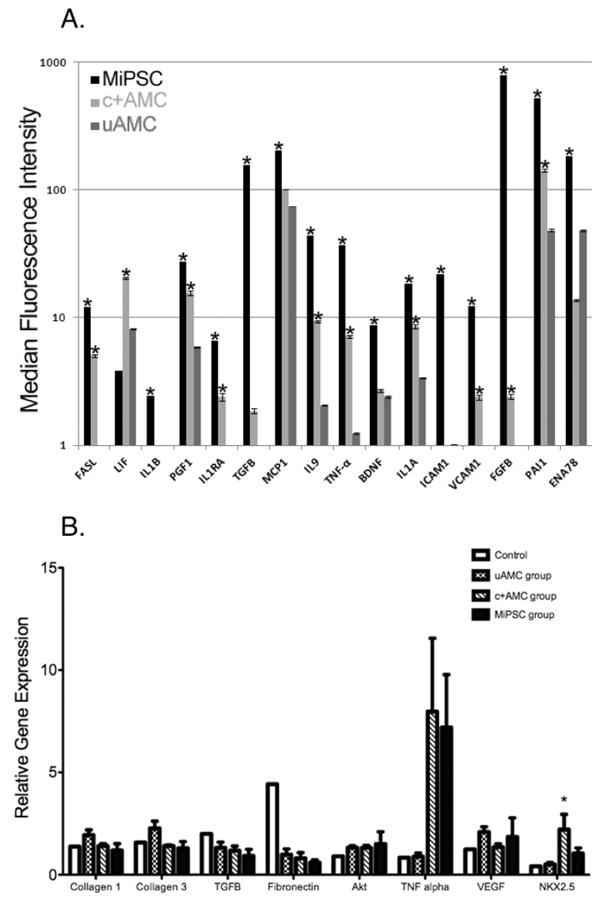
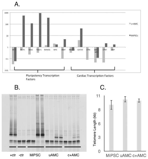
Rationale: The mechanism of functional restoration by stem cell therapy remains poorly understood. Novel manganese-enhanced MRI and bioluminescence reporter gene imaging were applied to follow myocardial viability and cell engraftment, respectively. Human-placenta-derived amniotic mesenchymal stem cells (AMCs) demonstrate unique immunoregulatory and precardiac properties. In this study, the restorative effects of 3 AMC-derived subpopulations were examined in a murine myocardial injury model: (1) unselected AMCs, (2) ckit(+)AMCs, and (3) AMC-derived induced pluripotent stem cells (MiPSCs).

Objective: To determine the differential restorative effects of the AMC-derived subpopulations in the murine myocardial injury model using multimodality imaging.

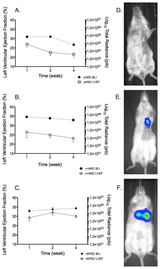
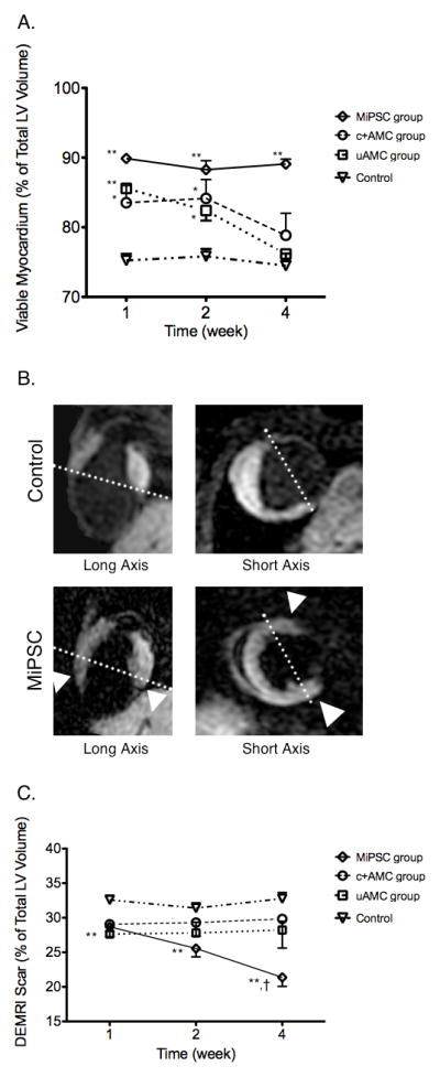
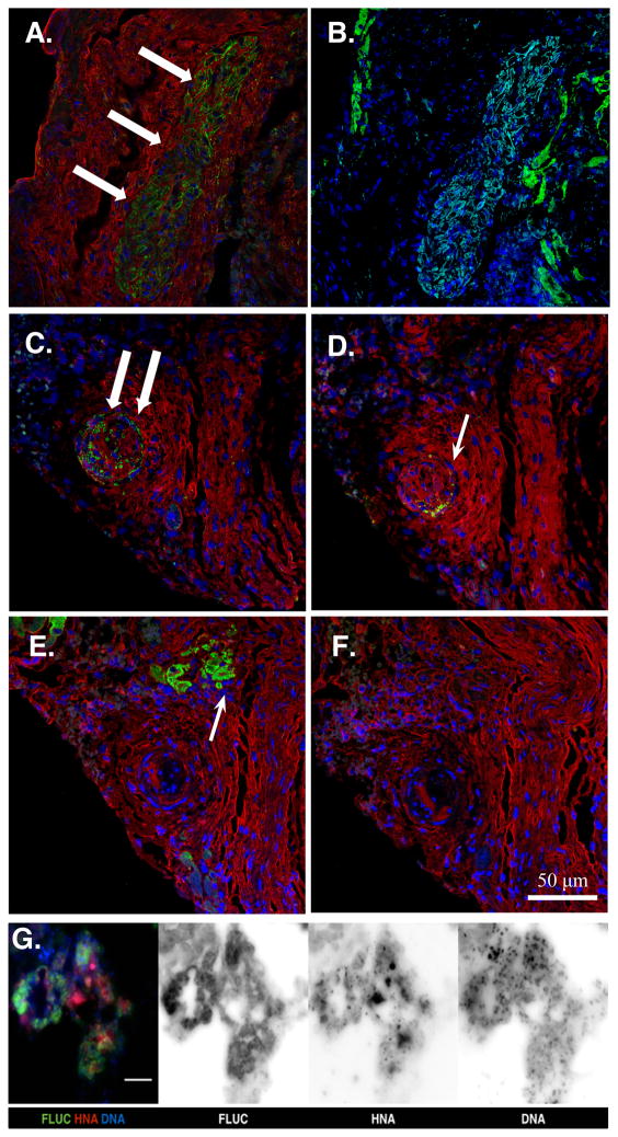
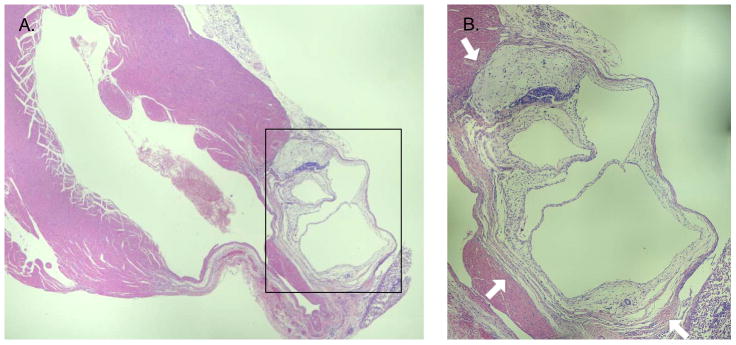
Methods and results: SCID (severe combined immunodeficiency) mice underwent left anterior descending artery ligation and were divided into 4 treatment arms: (1) normal saline control (n=14), (2) unselected AMCs (n=10), (3) ckit(+)AMCs (n=13), and (4) MiPSCs (n=11). Cardiac MRI assessed myocardial viability and left ventricular function, whereas bioluminescence imaging assessed stem cell engraftment during a 4-week period. Immunohistological labeling and reverse transcriptase polymerase chain reaction of the explanted myocardium were performed. The unselected AMC and ckit(+)AMC-treated mice demonstrated transient left ventricular functional improvement. However, the MiPSCs exhibited a significantly greater increase in left ventricular function compared with all the other groups during the entire 4-week period. Left ventricular functional improvement correlated with increased myocardial viability and sustained stem cell engraftment. The MiPSC-treated animals lacked any evidence of de novo cardiac differentiation.

Conclusion: The functional restoration seen in MiPSCs was characterized by increased myocardial viability and sustained engraftment without de novo cardiac differentiation, indicating salvage of the injured myocardium.

Keywords: magnetic resonance imaging; molecular imaging; multimodal imaging.


Figures

Figure 1
Figure 1. Effects of uAMCs, c+AMCs, and MiPSCs on LVEF. Stacks of short axis images acquired by CMR were analyzed offline to determine the LVEF for weeks 1, 2 and 4
(A) All groups treated with stem cells demonstrated improved LVEF initially compared to control. However, only the MiPSC group demonstrated sustained improvement through week 4. The c+AMC group demonstrated an intermediate restorative effect with significantly improved LVEF compared to control through weeks 1 and 2. The control group showed severely depressed LVEF that was unchanged throughout the study. (B) Short axis acquisitions are shown during end-diastole and end-systole at the mid-LV. The MiPSC treated mouse demonstrated increased contractility compared to control. (*p < 0.05, **p < 0.01 vs. control by unadjusted Student’s t-test.)
Figure 2
Figure 2. Effects of engraftment of uAMCs, c+AMCs and MiPSCs on LVEF demonstrate correlation between mean engraftment by BLI signal and LVEF by CMR
(A) Decreased stem cell engraftment and reduced LVEF in the uAMC group. (B) Decreased stem cell engraftment and reduced LVEF in the c+AMC group. (C) In contrast, sustained engraftment and improved LVEF in the MiPSC group throughout the 4 weeks. (D–F) BLI signal observed at week 4 for mice treated with uAMCs, c+AMCs, and MiPSCs respectively.
Figure 3
Figure 3. Effects of cell therapy as determined by MEMRI and DEMRI
(A) All groups treated with stem cells demonstrated increased viable myocardium by MEMRI, initially, compared to control. At week 4, only the MiPSC group demonstrated sustained increase in myocardial viability compared to control. The c+AMC and uAMC groups had decreasing viability that paralleled their reduced LVEF. (B) Representative MEMRI images from a control mouse and an MiPSC treated mouse are shown in the corresponding long- axis and short- axes. Increased myocardial viability was seen in the anterior and inferior walls of the MiPSC treated mouse (white arrowhead). (C) MiPSC group demonstrated significantly decreased myocardial scar by DEMRI while the uAMC and c+AMC groups demonstrated a trend towards decreased myocardial scar compared to control through the 4 week period. The difference between week 4 compared to week 1 was significant for the MiPSC group (†p = 0.01, *p < 0.05, **p < 0.01 vs. control.)
Figure 4
Figure 4. Immunohistology at the site of cell injection in MiPSC and c+AMC treated mice
(A) MiPSCs showed successful engraftment by the detection of human mitochondrial antibody (green, white arrows) at the site of cell injection. (B) Immunostaining with cardiac troponin T antibody (green) did not demonstrate any signal within the engrafted MiPSCs (light blue) and labeled only the native murine myocardium. (C) c+AMCs showed successful engraftment by human mitochondrial antibody (green, white arrow), (D) Persistent ckit+ expression was confirmed by ckit+ receptor antibody (green, white arrow). (E–F) However, immunostaining using cardiac troponin T (green, white arrow) and PECAM antibodies did not co-localize with the human mitochondrial or ckit+ stain demonstrating no evidence of cardiac or endothelial differentiation, respectively. Nuclear counterstain (blue) and F-actin (red) antibodies were used to visualize the cellular cytoskeleton. (G) Merged and monochrome images of the MiPSCs co-stained with anti-luciferase antibody (FLUC), human nuclear antibody (HNA), and Hoechst 33342 (DNA). Robust co-localization of the 3 immunostains confirms the origin of the BLI signal from the transplanted human stem cells.
Figure 5
Figure 5. Bright-field light micrograph of a teratoma at the peri-infarct cell injection site in a MiPSC treated mouse at 4 weeks
This micrograph was representative of teratomas observed in all explanted hearts of MiPSC treated mice. Three germ layers were identified confirming the presence of a teratoma. The teratoma formed from MiPSCs was loosely packed masses without intra-cavitary invasion typically seen with human ESCs. (A) 5X and (BB) 10X zoom.
Figure 6
Figure 6
(A) The 63-plex Luminex Immunoassay of human cytokines detected significant increase in the production of 15 cytokines in the supernatant of MiPSCs and of 10 cytokines in c+AMCs when compared to uAMCs (*p<0.05, Figure 6A). The significant production of cytokines in the MiPSCs was involved in anti-apoptosis (FASL, IL9, BDNF), anti-fibrosis (PAI1, TGF-B), pro-angiogenesis (VEGF, FGF-B, PGF1), and anti-inflammation (IL1A, IL1B, IL1RA, ICAM1, VCAM1, TNF-a, MCP1, ENA78). Similarly, c+AMCs showed increased production of 11 cytokines when compared to uAMCs : FASL, LIF, PGF1, IL1RA, TGF-B, IL9, TNF-a, IL1A, VCAM1, FGF-B, PAI1 (*p<0.05). (B) Effects of cell therapy on para-crine factor gene expression in the explanted myocardium as assayed by RT-PCR. Expression of fibrotic (collagen 1, collagen 3, fibronectin, Akt), apoptotic (TNF-alpha), angiogenic (VEGF), inflammatory (TGF-B), and cardiac specific (NKX2.5) genes were evaluated by RT-PCR at 4 weeks. Fibronectin, TNF-alpha, VEGF and NKX2.5 showed a trend towards differential gene expression in cell-based therapy treated groups compared to control. Only NKX2.5 gene expression in the c+AMC group demonstrated a significant increase compared to control (p = 0.04). *p < 0.05 vs. control.
Figure 7
Figure 7
(A) Stem cells demonstrated varying levels of pluripotency and cardiac lineage specification by RT-PCR. The relative gene expressions compared to uAMCs are shown. MiPSCs demonstrated significantly increased expression of early transcription factors compared to uAMCs, including OCT4, SOX2, TDGF1, NANOG, MYC, and EBAF. (B) Detection of telomerase activity in the MiPSCs by TRAP assay while uAMCs and c+AMCs did not exhibit increased telomerase activity (4,000 cells were used in each sample). (C) Mean telomere lengths in MiPSCs, uAMCs, and c+AMCs as measured by qPCR did not demonstrate any significant difference.

References

    1. Laflamme MA, Chen KY, Naumova AV, Muskheli V, Fugate JA, Dupras SK, Reinecke H, Xu C, Hassanipour M, Police S, O’Sullivan C, Collins L, Chen Y, Minami E, Gill EA, Ueno S, Yuan C, Gold J, Murry CE. Cardiomyocytes derived from human embryonic stem cells in pro-survival factors enhance function of infarcted rat hearts. Nat Biotechnol. 2007;25:1015–24. - PubMed
    1. Fernandes S, Naumova AV, Zhu WZ, Laflamme MA, Gold J, Murry CE. Human embryonic stem cell-derived cardiomyocytes engraft but do not alter cardiac remodeling after chronic infarction in rats. Journal of molecular and cellular cardiology. 2010;49:941–9. - PMC - PubMed
    1. Ratajczak MZ, Kucia M, Jadczyk T, Greco NJ, Wojakowski W, Tendera M, Ratajczak J. Pivotal role of paracrine effects in stem cell therapies in regenerative medicine: can we translate stem cell-secreted paracrine factors and microvesicles into better therapeutic strategies? Leukemia. 2012;26:1166–73. - PubMed
    1. Williams AR, Hare JM. Mesenchymal stem cells: biology, pathophysiology, translational findings, and therapeutic implications for cardiac disease. Circ Res. 2011;109:923–40. - PMC - PubMed
    1. Welt FG, Gallegos R, Connell J, Kajstura J, D’Amario D, Kwong RY, Coelho-Filho O, Shah R, Mitchell R, Leri A, Foley L, Anversa P, Pfeffer MA. Effect of cardiac stem cells on left-ventricular remodeling in a canine model of chronic myocardial infarction. Circ Heart Fail. 2013;6:99–106. - PubMed

Publication types

MeSH terms