Review
. 2015 Jun;1853(6):1253-71.
doi: 10.1016/j.bbamcr.2015.01.018. Epub 2015 Feb 2.

Emerging critical roles of Fe-S clusters in DNA replication and repair

Affiliations
Review

Emerging critical roles of Fe-S clusters in DNA replication and repair

Jill O Fuss et al. Biochim Biophys Acta. 2015 Jun.

Abstract

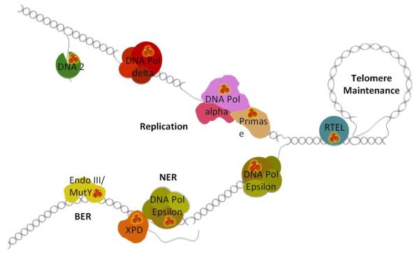
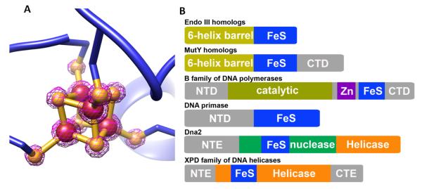
Fe-S clusters are partners in the origin of life that predate cells, acetyl-CoA metabolism, DNA, and the RNA world. The double helix solved the mystery of DNA replication by base pairing for accurate copying. Yet, for genome stability necessary to life, the double helix has equally important implications for damage repair. Here we examine striking advances that uncover Fe-S cluster roles both in copying the genetic sequence by DNA polymerases and in crucial repair processes for genome maintenance, as mutational defects cause cancer and degenerative disease. Moreover, we examine an exciting, controversial role for Fe-S clusters in a third element required for life - the long-range coordination and regulation of replication and repair events. By their ability to delocalize electrons over both Fe and S centers, Fe-S clusters have unbeatable features for protein conformational control and charge transfer via double-stranded DNA that may fundamentally transform our understanding of life, replication, and repair. This article is part of a Special Issue entitled: Fe/S proteins: Analysis, structure, function, biogenesis and diseases.

Keywords: Cancer and degenerative disease; DNA charge transfer communication; DNA repair; DNA replication; Fe–S cluster; Genome integrity.


Figures

Figure 1
Figure 1. The emerging roles of Fe-S cluster enzymes in DNA replication and repair
Replication: Fe-S clusters are critical elements of DNA primase, all replicative DNA polymerases (DNA pols α and δ shown), and the nuclease/helicase Dna2 (shown on lagging strand 5′ flaps). Nucleotide Excision Repair (NER): the 5′->3′ Fe-S cluster helicase XPD opens a single stranded bubble around duplex distorting DNA damage allowing excision of the damaged strand by endonucleases and the gap filling by DNA polymerase (DNA pol ε shown). Base Excision Repair (BER): glycosylases Endo III / MutY and their role in the discovery and removal of damaged and mispaired bases. Telomere Maintenance: the helicase RTEL is involved in the unwinding of telomeric D-loops that affects telomere length maintenance and HR in the region.
Figure 2
Figure 2. Structure of [4Fe-4S] clusters and their placement in DNA processing enzymes
A) [4Fe-4S] cluster with 2Fo-Fc map in contour 4ζ (PDB: 1WEI [251]). Fe and S are shown as brown and goldenrod spheres, respectively. B) Distinct schematic sequence architecture for Fe-S clusters in DNA replication and repair proteins. The distinct patterns of placement for the Fe-S clusters relative to catalytic domains suggest their sequence location along with their three-dimensional topology provide a potential means for differential DNA CT activities suitable to coordinate replication and repair pathways. In one of the simplest models, the C-terminal placement of the Fe-S clusters in glycosylases and polymerases might impact the DNA affinity and hence exchange rate versus processivity. The Dna2 helicase/nuclease and the XPD family helicases have Fe-S clusters inserted into catalytic domains suggesting a tight linkage between cluster and catalytic activities. The unique placement of the XPD family Fe-S cluster within the HD1 catalytic domain supports its role as a sensor for double helix disruption. These and other testable roles emerge from the sequence architectures and structures analyzed here.
Figure 3
Figure 3. Fe-S cluster domains and folds in DNA processing enzymes
Top: Ribbon diagrams of overall protein architecture (orange ribbon with surface representation) and placement of Fe-S cluster domains (colored in blue). Bottom: close-up view of Fe-S cluster domains. [4Fe-4S] cluster is shown as brown (Fe) and yellow (S) spheres. (A) crystal structure of XPD helicase from S. acidocaldarius (PDB: 3CRV [112]); (B) crystal structure of C-terminal domain (CTD) of catalytic subunit (blue) and B subunit (orange) complex of yeast DNA polymerase α (PDB: 3FLO [133]). Two Zn metals (gray) were bound to CTD. Zn-2 (CysB) binding site was later experimentally shown to be a Fe-S cluster, but Zn is bound in this structure; (C) structure of C-terminal regulatory domain of human DNA primase (PDB: 3L9Q [150]).
Figure 4
Figure 4. Important structure elements of EndoIII DNA interaction and MUTYH MAP mutations in the Fe-S domain
A) The overall structure of G. stearothermophilus Endonuclease III-DNA complex with [4Fe-4S] cluster (PDB:1P59 [80]). Fe-S cluster domain and DNA are shown in blue and green, respectively. B) Zoom in view of A show the interactions between Fe-S cluster domain and DNA form tightly H-bonding networks on Endo III-DNA complex (PDB:1P59 [80]). Conserved residue R144 forms H-bonding with residues C189, A191, and K190 that tightly hold Fe-S cluster domain with α-H helix together. That interaction positions conserved residue R148 to form H-bonding with DNA phosphate group and carbonyl group of G185 on α-J helix. And that positions residue R186 to form H-bonding with DNA phosphate and ribose groups. Interacting residues are shown in stick, H-bonding is shown in purple dot line, and Fe and S are shown in brown and yellow spheres, respectively. C) The interactions of human MUTYH associated polyposis (MAP) mutations with Fe-S cluster domain. MAP mutations R227W, R231C/H, V232F, and R195C (shown in cyan stick) form H-bonding network with Fe-S cluster domain. The similar interactions have been observed in EndoIII structure (Fig. 4B). Additionally, residue R295 forms H-bonding with E289 to fix the helix conformation for Fe-S cluster binding. Mutation of V232F can form steric clash with F82 that will destabilize the Fe-S cluster domain. (PDB:3N5N [108])
Figure 5
Figure 5. The XPD Fe-S cluster coordinates key structural elements and is important in human disease
A) Fe-S cluster stabilizes the local structure folding of Fe-S cluster domain in SaXPD (blue) (PDB:3CRV [112]). 55 residues in the Fe-S cluster domain becomes completely disordered without Fe-S cluster (cyan) (PDB:3CRW [112]). The Arch domain (orange) is also affected in the absence of Fe-S cluster (red). The disorder region is shown as green dot line. B) The impact of TTD mutation K84H on Fe-S cluster domain in SaXPD. Residue K84 forms hydrogen bonding (purple dot line) with one of the cysteine ligands of [4Fe-4S] cluster. [4Fe-4S] cluster was surrounded by many hydrophobic residues that control the solvent accessibility. The mutation of lysine to histidine can disrupt hydrogen-bonding interaction and impact the protein environment and redox potential of Fe-S cluster. The Fe-S cluster domain is shown in blue; residues K84 and hydrophobic residues are shown in stick.
Figure 6
Figure 6. DNA processing enzymes share common Fe-S cluster assembly machinery
A model for the Cytosolic Iron-Sulfur Cluster Assembly (CIA) in three steps. 1) The early steps of the mitochondrial Iron-Sulfur Cluster (ISC) machinery are essential for CIA. Many required proteins such as the cysteine desulfurase complex Nfs1-Isd11 and the Isu1 scaffold are not shown. An unknown sulfur-containing compound (X-S) is exported to the cytosol via ATM1, S-X potentially being a glutathione (GSH) coordinated [2Fe-2S] cluster. 2) X-S is transported to the Cfd1-Nbp35 scaffold complex that assembles the cytosolic [4Fe-4S] clusters. Monothiol glutaredoxins Grx3-Grx4 can transiently bind a [2Fe-2S] cluster and may help shuttle S-X to Cfd1-Nbp35. NTPase activity and electron transfer from Tah18-Dre2 is required for assembly in vivo. 3) IOP1 serves as a bridge between the scaffold and the CIA targeting complex of CIA1, CIA2B and MMS19. The targeting complex has been shown to have a long list of interactions with proteins known to have Fe-S clusters. The complex has not yet been shown to hold an Fe-S cluster, but may instead facilitate cluster handoff between IOP1 and target apoproteins.
Figure 7
Figure 7. The DNA mediated charge transport communication (DNA CTC) hypothesis as a method for protein-protein communication
A) DNA CT provides a means for Endo III and MutY to preferentially localize to damaged DNA due to a disruption in the DNA pi stack B) Proposed DNA CTC mechanism controls rapid switching between replicative polymerases during lagging strand synthesis C) Nucleotide Excision Repair (NER) DNA CTC between DNA polymerase and XPD to coordinate DNA synthesis, incision, and release of the damaged strand. D) DNA CTC communication between replicative polymerases at adjacent origins to control origin firing.

References

    1. Fontecave M. Iron-sulfur clusters: ever-expanding roles. Nat Chem Biol. 2006;2:171–174. doi:10.1038/nchembio0406-171. - PubMed
    1. Imlay JA. Iron-sulphur clusters and the problem with oxygen. Mol Microbiol. 2006;59:1073–1082. doi:10.1111/j.1365-2958.2006.05028.x. - PubMed
    1. Koonin EV, Martin W. On the origin of genomes and cells within inorganic compartments. Trends in Genetics. 2005;21:647–654. doi:10.1016/j.tig.2005.09.006. - PMC - PubMed
    1. Russell MJ, Martin W. The rocky roots of the acetyl-CoA pathway. Trends Biochem Sci. 2004;29:358–363. doi:10.1016/j.tibs.2004.05.007. - PubMed
    1. Imlay JA. Pathways of oxidative damage. Annu. Rev. Microbiol. 2003;57:395–418. doi:10.1146/annurev.micro.57.030502.090938. - PubMed

Publication types

LinkOut - more resources