. 2015 Apr;56(4):2348-57.
doi: 10.1167/iovs.14-15630.

Recruitment of Donor T Cells to the Eyes During Ocular GVHD in Recipients of MHC-Matched Allogeneic Hematopoietic Stem Cell Transplants

Affiliations

Recruitment of Donor T Cells to the Eyes During Ocular GVHD in Recipients of MHC-Matched Allogeneic Hematopoietic Stem Cell Transplants

Samantha Herretes et al. Invest Ophthalmol Vis Sci. 2015 Apr.

Abstract

Purpose: The primary objective of the present study was to identify the kinetics and origin of ocular infiltrating T cells in a preclinical model of graft-versus-host disease (GVHD) that induces eye tissue damage.

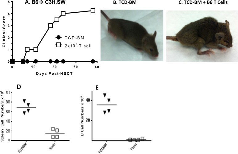
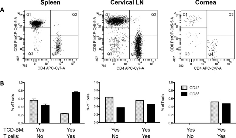
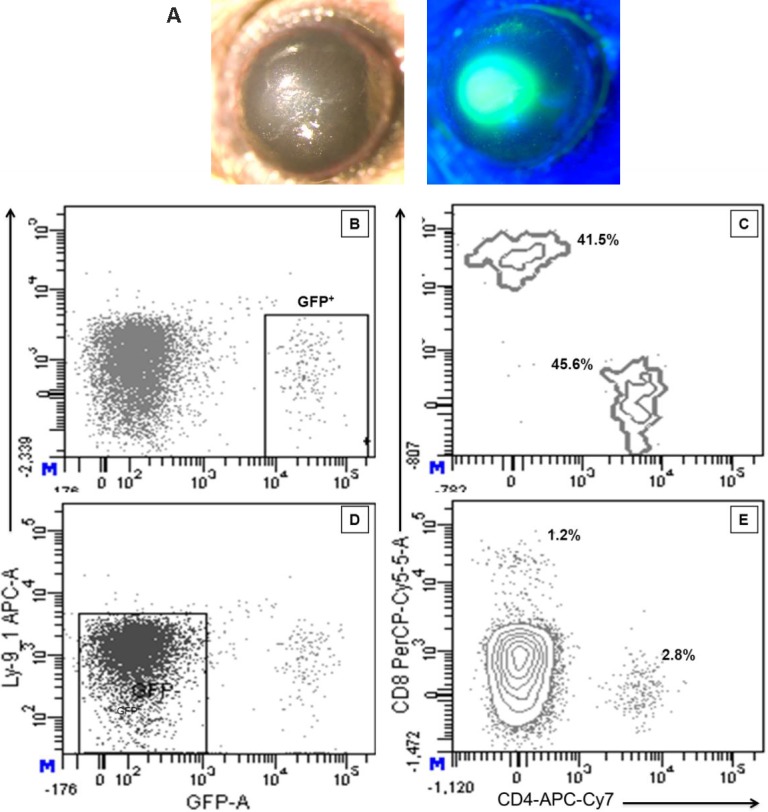
Methods: Graft-versus-host disease was induced using an major histocompatibility complex (MHC)-matched, minor histocompatibility-mismatched hematopoietic stem cell transplant (HSCT) model. This approach, which utilized congenic and EGFP-labeled donor populations, mimics a matched, clinically unrelated donor (MUD) cell transplant. Systemic and ocular GVHD were assessed at varying time points using clinical examination, intravital microscopy, immune phenotype via flow cytometric analyses, and immunohistochemical staining.

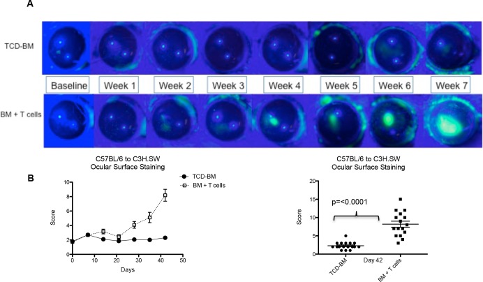
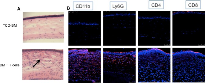
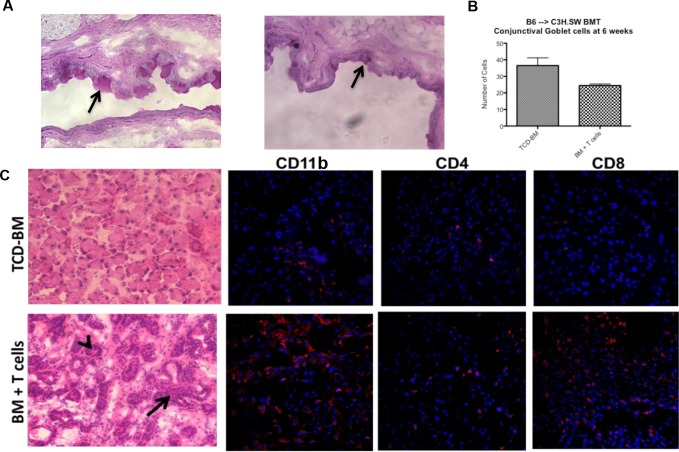
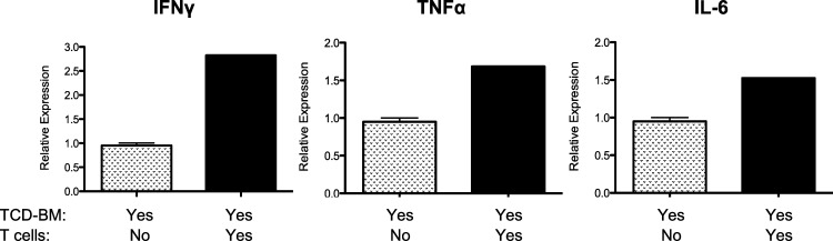
Results: Following transplant, we observed characteristic changes in GVHD-associated immune phenotype as well as clinical signs present in recipients post transplant. Notably, the kinetics of the systemic changes and the ocular damage paralleled what is observed clinically, including damage to the cornea as well as the conjunctiva and lacrimal gland. Importantly, the infiltrate contained predominantly donor CD4 as well as CD8 T cells with an activated phenotype and macrophages together with effector cytokines consistent with the presence of a TH1 alloreactive population.

Conclusions: Overall, the findings here unequivocally demonstrated that donor T cells compose part of the corneal and ocular adnexa infiltrate in animals undergoing ocular GVHD. In total, the results describe a novel and promising preclinical model characterized by both systemic and ocular changes as detected in significant numbers of patients undergoing GVHD following allo-HSCT, which can help facilitate dissecting the underlying immune mechanisms leading to damage associated with ocular GVHD.


Figures

Figure 1
Figure 1
Systemic GVHD in C3H.SW recipients post HSCT. (A) Mice that received T cells developed high systemic GVHD scoring. Photos representative of mice without (B) and with (C) GVHD characterized by weight loss, ruffled fur, and poor posture. Systemic GVHD was confirmed by low number of splenocytes (D) and B cells (E) in animals that received TCD-BM + T cells.
Figure 2
Figure 2
Ocular surface assessment post HSCT: clinical changes in the corneas of HSCT recipients. (A) Clinical photos through 7 weeks demonstrating progression of corneal staining and development of ulcers in the group that received transplantation with TCD-BM + T cells. (B) Quantification of corneal fluorescein staining throughout the study and at day 42 after transplantation.
Figure 3
Figure 3
Pathologic changes in the corneas of allogeneic HSCT recipients. (A) Photographs (H&E stained, 7-μm sections, ×20) of the central corneas of mice receiving B6 TCD-BM only or together with B6 T cells. Dense cellular infiltrates (black arrow) are observed in the eyes of animals with systemic GVHD. (B) Immunofluorescent microscopy. Photographs of 7-μm sections at ×40 stained with CD11b (macrophages), Ly6G (neutrophils), CD4, CD8 (T cells), mAbs (red), and DAPI (blue). Cellular infiltrates in animals with GVHD consist of T cells, macrophages, and neutrophils.
Figure 4
Figure 4
Involvement of the ocular adnexa in mice with GVHD. (A) Photographs (PAS-stained, 7-μm sections, ×20) of the central superior conjunctiva of mice receiving B6 TCD-BM only or together with B6 T cells, where goblet cells stain dark pink (black arrow). Mice with GVHD exhibited thickening and irregularity of the basal membrane, as well as atrophy and reduced number of goblet cells (B). (C) Photographs of lacrimal gland histology from mice receiving TCD-BM only or together with T cells. Sections are 7 μm thick and stained with H&E at ×20 magnification. Periductal fibrosis, dense cellular infiltrates (black arrow), and stasis of secretions in ducts (arrowheads) are observed in lacrimal glands of animals with systemic GVHD.
Figure 5
Figure 5
Identification of donor T-cell populations in recipient corneas of mice with GVHD. Mice received BM-TCD from B6-CD45.1 donors + T cells from B6-EGFP donors. Top: Donor EGFP+ T cells were identified in vivo in the recipient corneas using fluorescent stereomicroscopy by 2 to 3 weeks post transplant (top middle and right). Clinical examination of the ocular surface (bottom) demonstrated the development of corneal ulcers approximately 4 to 6 weeks post transplant (bottom right, arrow). Data presented are from individual transplant recipient. T-cell infiltrates preceding clinical evidence of ocular surface disease were observed in seven mice in two independent experiments.
Figure 6
Figure 6
CD4/CD8 ratio differs dependent on the “target tissue.” Mice received BM-TCD from B6-CD45.1 donors + T cells from B6-EGFP donors. (A) Spleens of mice that received TCD-BM + T cells displayed the characteristic GVHD phenotype, with increased number of CD8+ T cells compared to CD4+ cells, and a decreased CD4+/CD8+ ratio. Data represent analysis of cells gated on EGFP+ cells. Quadrants represent Q1, donor GFP+CD8+ (PerCP-Cy5-5A); Q3, CD4 CD8; Q4, donor GFP+CD4+(APC-Cy7-A). Interestingly, ocular compartments such as cornea and draining cervical lymph nodes showed equal or even higher amounts of CD4+ cells compared to CD8+ T cells, creating a higher CD4/8 ratio. (B) Spleen: Data represent three to five individual spleens/group from one of two independent experiments. Cervical lymph nodes and cornea: Data represent pool of three to five mice/group from one of two independent experiments. Data are presented as fraction of total lymphocytes in each compartment.
Figure 7
Figure 7
Subset analyses of T-cell infiltrate in the corneas of recipients with GVHD. Mice that received B6-CXCR6−/− EGFP developed clinical ocular changes characterized by ulceration (A). Flow cytometric analysis of corneal cell suspensions 5 weeks post transplant demonstrated the presence of a EGFP+Ly9.1 population (B), which contained both CD4 and CD8 lymphoid subsets (C), confirming the presence of donor T cells in the recipient corneas. The ocular infiltrate also contained GFP cells (D) that could represent T cells (E) derived from either transplanted donor progenitor cells or the recipient. Results represent cells obtained from 10 pooled corneas (five transplant recipients).
Figure 8
Figure 8
Inflammatory cytokines are elevated in the cornea of mice with ocular GVHD. RNA was prepared from corneal lysates obtained from C3H.SW recipients of B6 TCD-BM alone or B6 T cell–replete transplants (Table, line 1), and IL-6 mRNA was assessed by RT-PCR (see Methods). Results represent the mean of duplicate samples analyzed from pooled cornea extracts (n = 4 corneas from two recipients) 4 to 5 weeks post transplant.

References

    1. Li HW,, Sykes M.Emerging concepts in haematopoietic cell transplantation. Nat Rev Immunol. 2012; 12: 403–416. - PMC - PubMed
    1. Martin PJ,, Inamoto Y,, Carpenter PA,, Lee SJ,, Flowers MED.Treatment of chronic graft-versus-host disease: past, present and future. Korean J Hematol. 2011; 46: 153–163. - PMC - PubMed
    1. Arora M,, Klein JP,, Weisdorf DJ,, Hassebroek A,, Flowers ME,, Cutler CS,, et al. Chronic GVHD risk score: a Center for International Blood and Marrow Transplant Research analysis. Blood. 2011; 117: 6714–6720. - PMC - PubMed
    1. Blazar BR,, Murphy WJ,, Abedi M.Advances in graft-versus-host disease biology and therapy. Nat Rev Immunol. 2012; 12: 443–458. - PMC - PubMed
    1. Ferrara JL,, Levine JE,, Reddy P,, Holler E.Graft-versus-host disease. Lancet. 2009; 373: 1550–1561. - PMC - PubMed

Publication types