. 2015 Feb 5;56(2):1374-86.
doi: 10.1167/iovs.14-15196.

Identification of p58IPK as a novel neuroprotective factor for retinal neurons

Affiliations

Identification of p58IPK as a novel neuroprotective factor for retinal neurons

Evgenii Boriushkin et al. Invest Ophthalmol Vis Sci. .

Abstract

Purpose: Endoplasmic reticulum (ER)-resident chaperone protein p58(IPK) plays a vital role in regulation of protein folding and biosynthesis. The goal of this study was to examine the role of p58(IPK) in retinal neuronal cells under normal and stressed conditions.

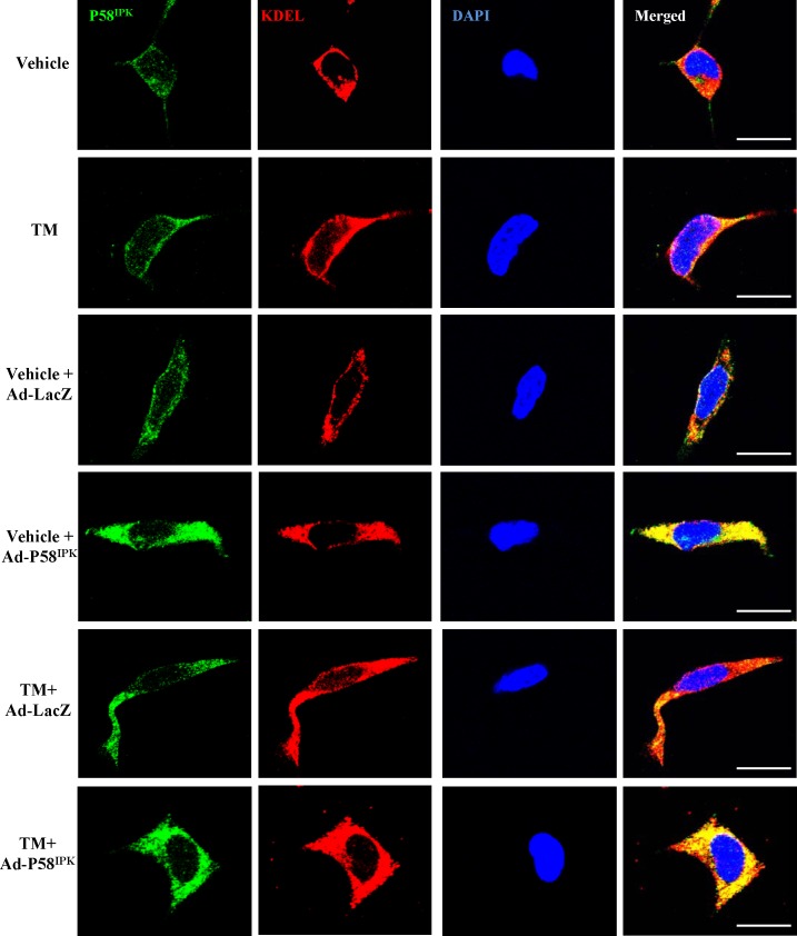
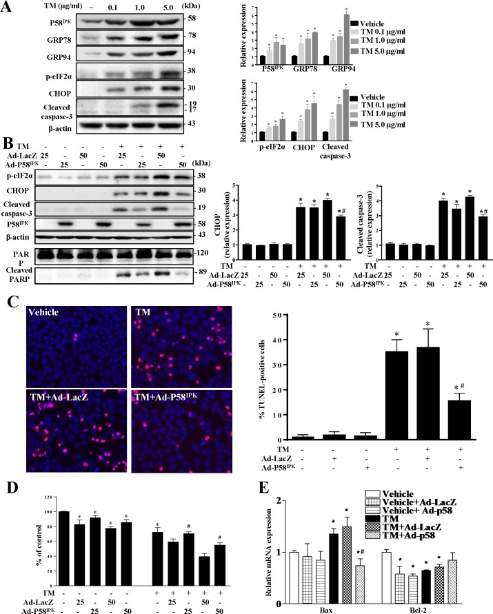
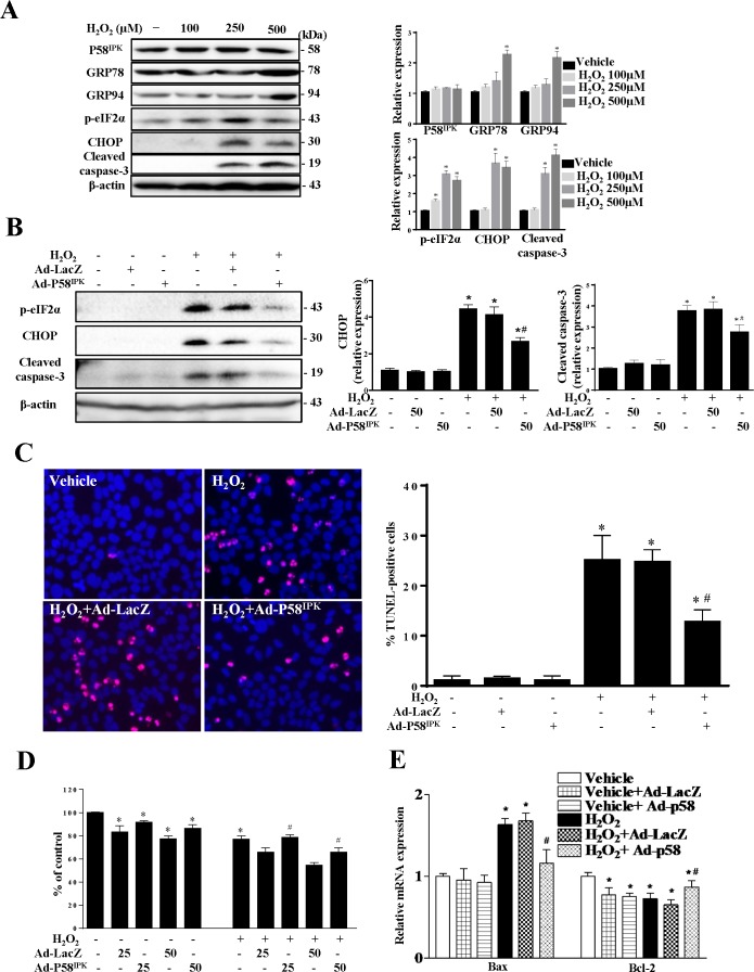
Methods: Retinal expression of p58(IPK), retinal morphology, apoptosis, ER stress, and apoptotic gene expression were examined in p58(IPK) knockout (KO) and/or wild-type (WT) mice with or without intravitreal injection of N-methyl-D-aspartic acid (NMDA). In in vitro experiments, differentiated R28 retinal neuronal cells transduced with adenovirus encoding p58(IPK) (Ad-p58(IPK)) or control virus (Ad-LacZ) were exposed to tunicamycin (TM) or hydrogen peroxide (H2O2). Levels of ER stress, apoptosis, and cell survival were evaluated.

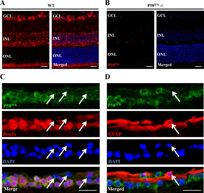
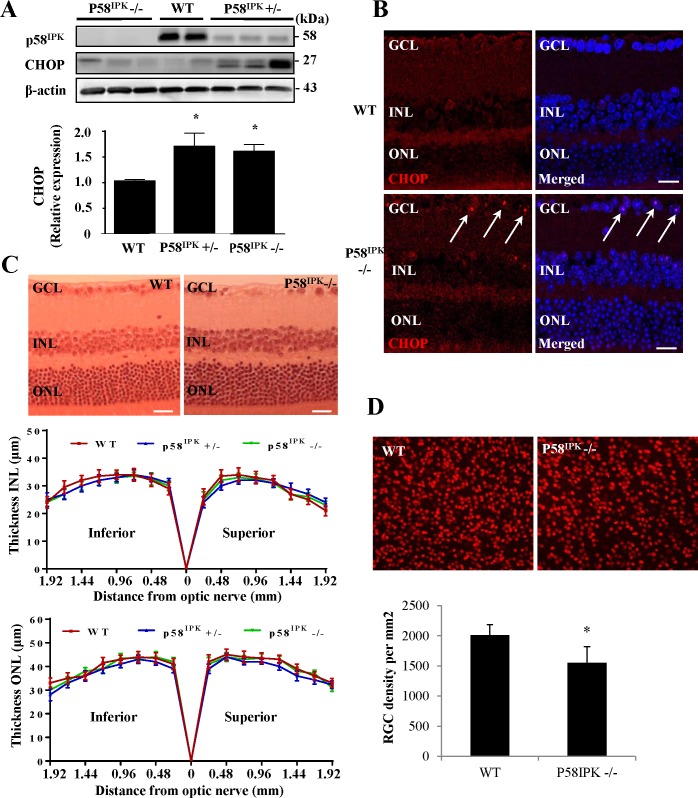
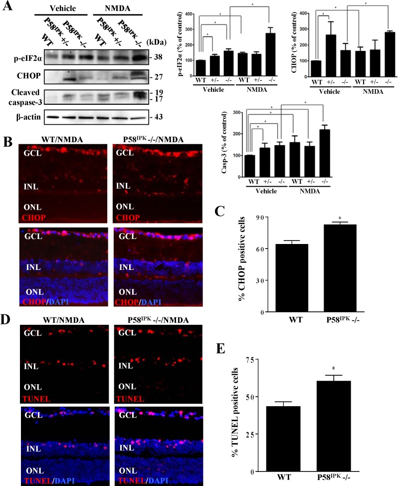
Results: Chaperone protein p58(IPK) is expressed predominantly in retinal ganglion cells (RGC), inner retinal neurons, and the photoreceptor inner segments. Mice lacking p58(IPK) exhibited increased CHOP expression and loss of RGCs with aging (8-10 months). Intravitreal injection of NMDA induced retinal ER stress and increased p58(IPK) expression in WT mice; this resulted in greater ER stress and enhanced RGC apoptosis in p58(IPK) KO mice. In cultured R28 cells, overexpression of p58(IPK) significantly reduced eIF2α phosphorylation, decreased CHOP expression, and alleviated the activation of caspase-3 and PARP. Overexpression of p58(IPK) also protected against oxidative and ER stress-induced cell apoptosis. Furthermore, p58(IPK) downregulated the proapoptotic gene Bax and upregulated the antiapoptotic gene Bcl-2 expression in stressed R28 cells.

Conclusions: Our study has demonstrated a protective role of p58(IPK) in retinal neurons, which may act in part through a mechanism involving modulation of ER homeostasis and apoptosis, particularly under conditions of cellular stresses.

Keywords: chaperone; endoplasmic reticulum stress; neuronal cells; p58IPK.


Figures

Figure 1
Figure 1
Protein p58IPK is highly expressed in RGCs in mice. (A). Immunostaining shows immunoreactivity of p58IPK in the retinal GCL, INL, and the inner segments of photoreceptors in the ONL in WT mice. Staining for p58IPK is shown in red and nuclei are stained in blue with DAPI. (B) Immunostaining for p58IPK in retinal sections from p58IPK KO mice. Nuclei are stained in blue with DAPI. (C) Representative images show that Brn3a (red)-positive ganglion cells exhibit high levels of p58IPK (green) in the GCL of WT mouse retina. White arrows indicate low p58IPK immunoreactivity in Brn3a-negative cells in GCL. Nuclei stained in blue with DAPI. (D). Representative images show that GFAP (red)-positive astrocytes demonstrate low levels of p58IPK (white arrows) compared with ganglion cells in GCL. Nuclei are stained in blue with DAPI. All results represent at least three mice in each group. Scale bar: 20 μm.
Figure 2
Figure 2
Loss of RGCs and increased CHOP expression in p58IPK KO mice. (A) Immunoblotting of CHOP and p58IPK in retinas from homozygous (p58IPK−/−) and heterozygous (p58IPK ±) p58IPK KO and WT mice. Protein levels of CHOP were quantified by densitometry (means ± SD, n = 4). *P < 0.05 versus WT mice. (B) Immunostaining of CHOP (red) in retinas from WT and p58IPK−/− mice. Nuclei were stained in blue with DAPI. Left panels: CHOP staining (red). Right panels: Merged images of CHOP (red) with DAPI (blue) staining. White arrows indicate increased expression of CHOP in ganglion cells. Results represent at least three mice in each group. (C) Upper panel: Representative images of retinal sections (1.0 mm from the optic nerve) from 8- to 10-month-old female WT and p58IPK−/− mice stained with hematoxylin-eosin. Lower panels: Quantification of INL and ONL thickness. Data are expressed as means ± SD, n = 6. (D) Upper panel: Representative images of fluorescent microphotographs of flat-mounted retinas from 8- to 10-month-old female WT and p58IPK−/− mice. Retinal ganglion cells are shown by their immunoreactivity of Brn3a in red. Lower panel: Quantification of the number of retinal ganglion cells. Data are expressed as means ± SD, n = 4. *P < 0.05 versus WT mice.
Figure 3
Figure 3
Deletion of p58IPK increases phosphorylation of eIF2α, CHOP expression and ganglion cell apoptosis after NMDA treatment. Four-month-old female WT and p58IPK−/− mice received an intravitreal injection of NMDA (20 nmol/eye) in one eye and vehicle in the contralateral eye. (A) Immunoblotting of protein extract from retinas after 8 hours postinjection show increased phosphorylation of eIF2α, expression of CHOP and activation of caspase-3 in p58IPK−/− compared with WT mice. Protein levels of p-eIF2α, CHOP, and cleaved caspase-3 were quantified by densitometry (means ± SD, n = 4). *P < 0.01. (B). Representative images of immunostaining of mouse retina for CHOP (red) in WT and p58IPK−/− mice after NMDA treatment. Nuclei were stained blue with DAPI. Upper panels: CHOP staining (red). Lower panels: Merged images of CHOP (red) and DAPI (blue) staining. (C) Quantification of the number of CHOP positive cells in GCL in retinal sections from 4 p58IPK−/− and 4 WT mice. Data are means ± SD. *P < 0.01 versus WT mice. (D) Representative images of TUNEL staining of mouse retina in p58IPK−/− and WT mice. Upper panels: TUNEL staining (red). Lower panels: merged images of TUNEL (red) and DAPI (blue) staining. (E) Quantification of the number of total and TUNEL positive cells in GCL in retinal sections from 4 p58IPK−/− and four WT mice. Data are expressed as means ± SD. *P < 0.01 versus WT mice.
Figure 4
Figure 4
Protein p58IPK primarily localizes in the ER in R28 cells. Representative images show expression and localization of p58IPK (green) in R28 cells. We used KDEL (red) as a marker of the ER and nuclei were stained by DAPI in blue. After treatment with TM, expression of p58IPK increased and colocalized with KDEL. After viral transduction, exogenous p58IPK also colocalized with KDEL in the vehicle and TM-treated cells. Scale bar: 20 μm.
Figure 5
Figure 5
Overexpression of p58IPK reduces ER stress and apoptosis in R28 cells after treatment with TM. (A) We treated R28 cells with 0.1 to 5 μg/mL of tunicamycin (TM) for 24 hours. Left panel: Immunoblots showing increased expression of GRP78, GRP94, CHOP, phosphorylation of eIF2α and activation of caspase-3 in a dose-dependent manner. Right panels: quantification of protein expression by densitometry (means ± SD, n = 4). *P < 0.001 versus vehicle. (B) R28 cells were transduced with 25 or 50 MOI of adenovirus for 24 hours, followed by treatment with 1.0 μg/mL TM for an additional 24 hours. Expression of ER stress markers and apoptotic proteins was determined by immunoblotting. Left panel: Shows representative images of blots. Right panels: Show the quantification results by densitometry (means ± SD, n = 4). *P < 0.001 versus corresponding vehicle control. #P < 0.05 versus corresponding Ad-LacZ group with TM treatment. (C) R28 cells transduced with 50 MOI of Ad-p58IPK or Ad-LacZ were treated with 1.0 μg/mL TM for 24 hours. Left panels: Representative images of TUNEL staining. TUNEL-positive nuclei of cells are pink (colocalization of TUNEL, red staining, with nuclear staining, blue with DAPI). Right panel: Quantification of TUNEL-positive cells in 6 to 9 frames/group. Results were expressed as means ± SD from four independent experiments. *P < 0.001 versus to vehicle without adenovirus. #P < 0.01 versus Ad-LacZ + TM. (D) Cell viability measured by MTT assay. Results are means ± SD of four independent experiments. *P < 0.001 versus vehicle without adenovirus, #P < 0.05 versus the corresponding Ad-LacZ group with TM treatment. (E) Results of qRT-PCR showing relative gene expression levels for Bax and Bcl-2. Results are means ± SD of four independent experiments. *P < 0.01 versus vehicle without adenovirus, #P < 0.05 versus Ad-LacZ + TM.
Figure 6
Figure 6
Overexpression of p58IPK attenuates ROS-induced ER stress and apoptosis. (A) R28 cells were treated with H2O2 for 24 hours. Left panel: Immunoblots showing increased expression of GRP78 and CHOP, phosphorylation of eIF2α and activation of caspase-3 by H2O2 in a dose-dependent manner. Right panels: The protein expression was semiquantified by densitometry (mean ± SD, n = 4). *P < 0.01 versus vehicle. (B) R28 cells were transduced with Ad-p58IPK and Ad-LacZ for 24 hours, followed by treatment with 500 μM H2O2 for 8 hours. Left panel: Immunoblotting demonstrating that overexpression of p58IPK decreases expression of CHOP, phosphorylation of eIF2α and activation of caspase-3 in H2O2-treated cells. Right panels: quantification of protein expression by densitometry (mean ± SD, n = 4). *P < 0.001 versus vehicle. #P < 0.01 versus Ad-LacZ + H2O2. (C) Left panels: Representative images of TUNEL staining in adenoviral transduced cells after treatment with 500 μM H2O2 for 8 hours. Right panel: quantification of TUNEL-positive cells. Results are means ± SD of 6 to 9 frames/group from four independent experiments. *P < 0.001 versus vehicle. #P < 0.01 versus Ad-LacZ + H2O2. (D) Cell viability measured by MTT assay in cells after H2O2 treatment for 8 hours. Results are means ± SD of four independent experiments. *P < 0.001 versus vehicle. #P < 0.05 versus corresponding Ad-LacZ + H2O2 group. (E) Results of qRT-PCR showing relative gene expression levels for Bax and Bcl-2. Results are mean ± SD of four independent experiments. *P < 0.01 versus vehicle. #P < 0.05 versus Ad-LacZ + H2O2.

References

    1. Xu C, Bailly-Maitre B, Reed JC. Endoplasmic reticulum stress: cell life and death decisions. J Clin Invest. 2005; 115: 2656–2664. - PMC - PubMed
    1. Ron D, Walter P. Signal integration in the endoplasmic reticulum unfolded protein response. Nat Rev Cell Biol. 2007; 8: 519–529. - PubMed
    1. Rutkowski DT, Kang SW, Goodman AG, et al. The role of p58IPK in protecting the stressed endoplasmic reticulum. Mol Biol Cell. 2007; 18: 3681–3691. - PMC - PubMed
    1. Tao J, Wu Y, Ron D, Sha B. Preliminary x-ray crystallographic studies of mouse UPR responsive protein P58(IPK) TPR fragment. Acta Crystallogr Sect F Struct Biol Cryst Commun. 2008; 64: 108–110. - PMC - PubMed
    1. Tao J, Sha B. Structural insight into the protective role of P58(IPK) during unfolded protein response. Methods Enzymol. 2011; 490: 259–270. - PubMed

Publication types

MeSH terms