. 2015 Feb 6:5:8287.
doi: 10.1038/srep08287.

Natural Green coating inhibits adhesion of clinically important bacteria

Affiliations

Natural Green coating inhibits adhesion of clinically important bacteria

Danielle S Trentin et al. Sci Rep. .

Abstract

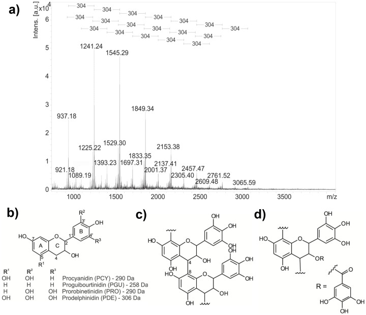
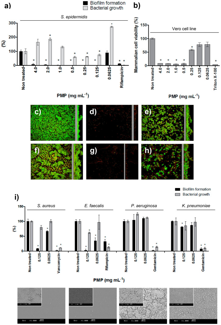
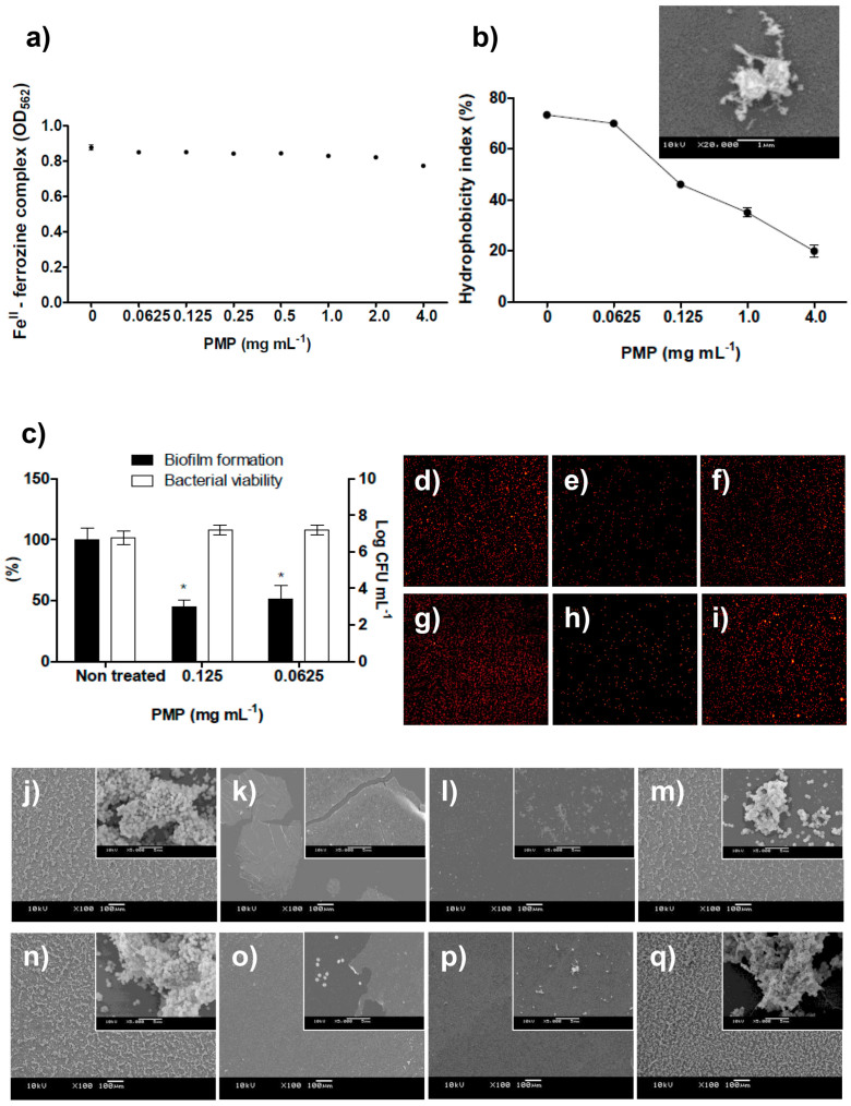
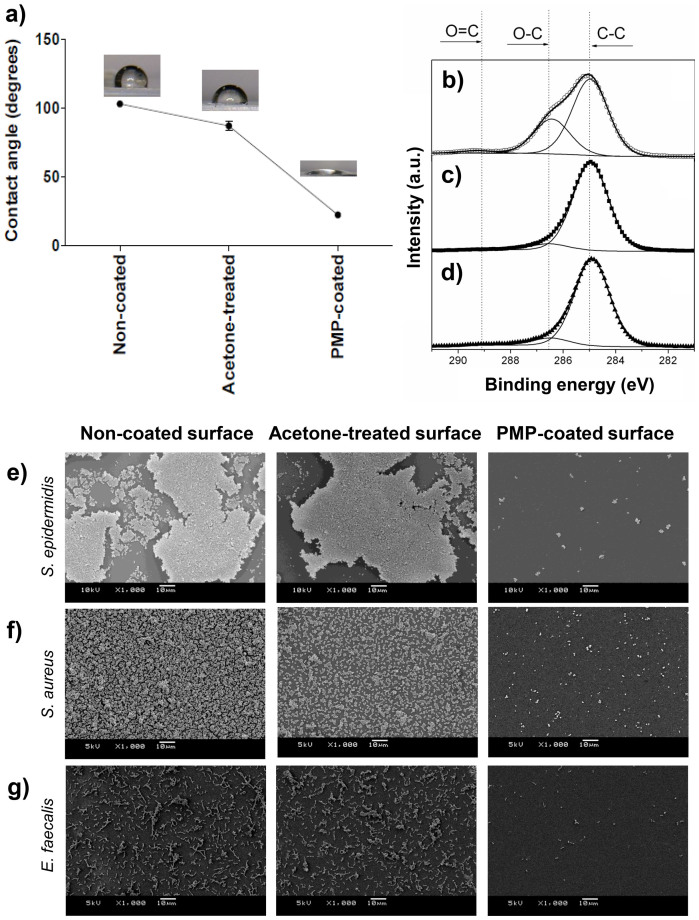
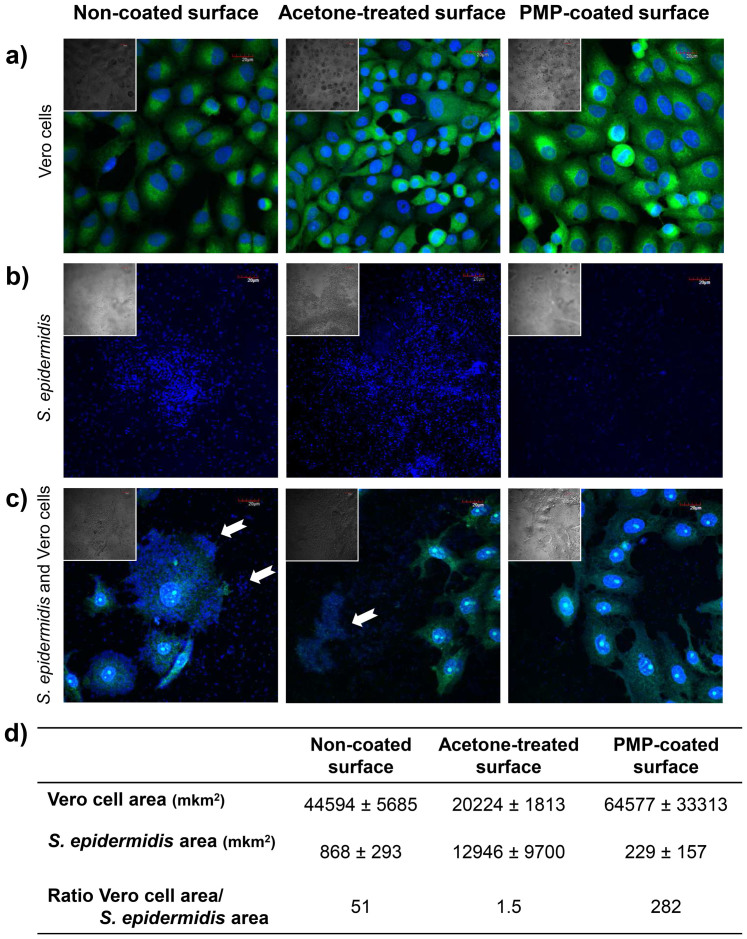
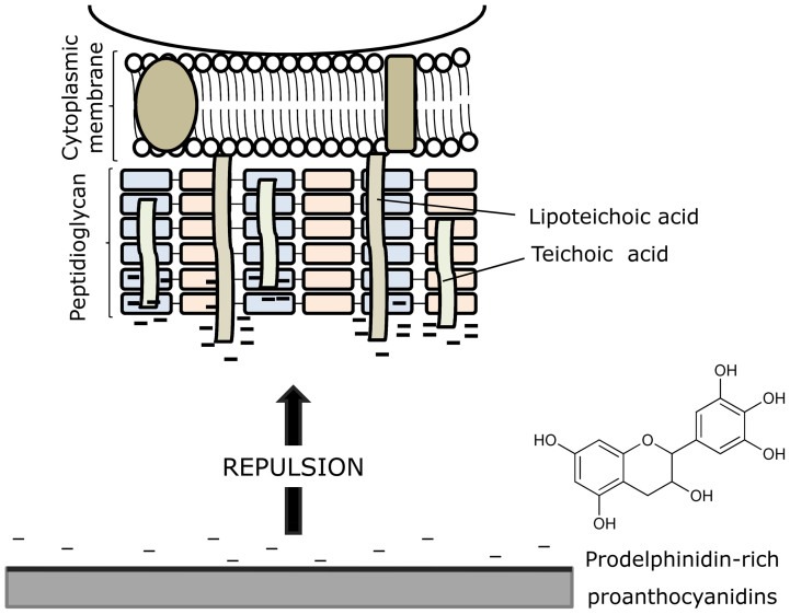
Despite many advances, biomaterial-associated infections continue to be a major clinical problem. In order to minimize bacterial adhesion, material surface modifications are currently being investigated and natural products possess large potential for the design of innovative surface coatings. We report the bioguided phytochemical investigation of Pityrocarpa moniliformis and the characterization of tannins by mass spectrometry. It was demonstrated that B-type linked proanthocyanidins-coated surfaces, here termed Green coatings, reduced Gram-positive bacterial adhesion and supported mammalian cell spreading. The proposed mechanism of bacterial attachment inhibition is based on electrostatic repulsion, high hydrophilicity and the steric hindrance provided by the coating that blocks bacterium-substratum interactions. This work shows the applicability of a prototype Green-coated surface that aims to promote necessary mammalian tissue compatibility, while reducing bacterial colonization.


Figures

Figure 1
Figure 1. Proanthocyanidins from P. moniliformis leaves.
(a) Mass spectra (positive ionization mode) of PMP, highlighting the consecutive mass differences of 304 Da in the polymeric series; (b) chemical structures of proanthocyanidin repeating units: procyanidin (PCY), proguibourtinidin (PGU), prorobinetinidin (PRO) and prodelphinidin (PDE); (c) typical linear proanthocyanidins possessing B-type linkage and (d) the proanthocyanidin with galloyl unit (m/z 785 [M + Na]+) identified in series C from PMP.
Figure 2
Figure 2. The effect of different concentrations of PMP on bacterial biofilm formation and growth, and on viability of epithelial mammalian cells.
(a) Dose-response curve of PMP tested against S. epidermidis biofilm formation and bacterial growth. (b) Dose-response curve of PMP tested upon mammalian cells viability. The biofilm topography of non-treated S. epidermidis on (c) polystyrene and on (f) glass surfaces; the exposition of S. epidermidis to 0.125 mg mL−1 of PMP (d and g, respectively to polystyrene and glass) and the biofilm formation when cells were exposed to 0.0625 mg mL−1 (e and h, respectively to polystyrene and glass). CLSM methodology can be found in Supplementary Methods. (i) Effect of PMP on other bacterial species involved in implant infections. SEM images show bacteria after exposure to 0.125 mg mL−1 and inserts present the respective non-treated control (bars indicate 10 μm). * represents statistical difference (p < 0.05) between treated and non-treated samples when analyzed by Student's t-test.
Figure 3
Figure 3
(a) The concentration of ferrozine-FeII complex was not decreased in the presence of PMP, indicating that these proanthocyanidins are not strong FeII chelators (the standard curve established to determine the FeII concentration to be used in the ferrozine assay and the curve of positive-chelator 2,2-bipyridyl can be found in Supplementary Fig. S10). (b) The dose-dependent decreasing of S. epidermidis surface hydrophobicity index (HBPI); values of HPBI greater than 70% indicated hydrophobic bacterial surface (non-treated or treated with PMP at 0.0625 mg mL−1); less than 70% indicated hydrophilic bacterial surface. SEM image shows that surface of S. epidermidis cells become covered by amorphous material in solutions containing PMP. (c) S. epidermidis partially recovered the ability to form biofilm and remained viable after exposure to proanthocyanidins for 24 h with three subsequent washes using sterile 0.9% NaCl. * represents statistical difference (p < 0.05) between treated and non-treated samples when analyzed by Student's t-test. (d–i) Fluorescence microscopy images (10x magnification) demonstrated that (d and g) non-treated microspheres and (f and i) microspheres treated with 0.0625 mg mL−1 of PMP attach to Permanox and to glass surfaces, respectively, while attachment was inhibited for spheres exposed to 0.125 mg mL−1 of PMP (e and h, respectively for Permanox and glass surfaces), similarly as observed for S. epidermidis cells treated with PMP. similarly as observed for S. epidermidis cells treated with PMP. (j–q) SEM images of S. epidermidis treated with PMP. Untreated S. epidermidis displayed several microcolonies on (j) Permanox and (n) on glass surfaces. When S. epidermidis was exposed to 4.0 mg mL−1 of PMP, there was no sign of attached cells while some regions of (k) Permanox and (o) glass substrates, and PMP spontaneously adhered on hydrophobic and hydrophilic substrates (note the insert showing that single cells adhered where there is no PMP film). Inhibition of biofilm formation at 0.125 mg mL−1 of PMP is observed on (l) Permanox and on (p) glass surfaces. The ability of S. epidermidis to form protective biofilm is not inhibited by 0.0625 mg mL−1 of PMP neither on (m) Permanox nor on (q) glass substrates. Bars represent 100 μm and in the inserts, 5 μm. All of these methodologies can be found in Supplementary Methods.
Figure 4
Figure 4. Green-coated surface analyses.
(a) WCA of non-coated, acetone-treated and PMP coated-surfaces. (b) XPS analyses of PMP coated-surfaces, (c) acetone-treated and (d) non-coated surfaces. SEM images of adhesion and biofilm formation by (e) S. epidermidis, (f) S. aureus and (g) E. faecalis on non-coated surface, acetone-treated surface and PMP-coated surface, respectively. Bars indicate 10 μm.
Figure 5
Figure 5. Co-culture of bacteria and mammalian cells using the perioperative bacterial model.
(a) Adhesion and spreading of mammalian Vero cells, (b) adhesion of S. epidermidis, (c) co-culture of S. epidermidis and mammalian Vero cells on non-coated, acetone-treated and PMP-coated surfaces, respectively; and (d) quantitative data of co-culture experiments as measurements of mammalian and bacterial cell area. Samples were imaged by 60x CLSM (FLUTAX-2 labels microtubules and DAPI stains DNA) and by differential interference contrast (DIC – in the inserts). White arrows indicate bacterial cell clusters. Bars in the images indicate 20 μm.
Figure 6
Figure 6. Mechanism proposed for inhibition of bacterial adhesion and biofilm formation by PMP-coated surfaces.
It is based on repulsive forces between the anionic S. epidermidis surface and the negatively charged material surface after coating with proanthocyanidins.

References

    1. Bazaka K., Jacob M. V., Crawford R. J. & Ivanova E. P. Efficient surface modification of biomaterial to prevent biofilm formation and the attachment of microorganisms. Appl. Microbiol. Biotechnol. 95, 299–311 (2012). - PubMed
    1. Langer R. & Tirrell D. A. Designing materials for biology and medicine. Nature 428, 487–492 (2004). - PubMed
    1. Holzapfel B. M. et al. How smart do biomaterials need to be? A translational science and clinical point of view. Adv. Drug Deliv. Rev. 65, 581–603 (2013). - PubMed
    1. Busscher H. J. et al. Biomaterial-associated infection: locating the finish line in the race for the surface. Sci. Transl. Med. 4, 153rv10 (2012). - PubMed
    1. Bjarnsholt T., Ciofu O., Molin S., Givskov M. & Høiby N. Applying insights from biofilm biology to drug development - can a new approach be developed? Nat. Rev. Drug Discov. 12, 791–808 (2013). - PubMed

Publication types

MeSH terms

LinkOut - more resources