. 2015 Mar;77(3):156-65.
doi: 10.1016/j.jdermsci.2015.01.001. Epub 2015 Jan 14.

Gene expression profiling in pachyonychia congenita skin

Affiliations

Gene expression profiling in pachyonychia congenita skin

Yu-An Cao et al. J Dermatol Sci. 2015 Mar.

Abstract

Background: Pachyonychia congenita (PC) is a skin disorder resulting from mutations in keratin (K) proteins including K6a, K6b, K16, and K17. One of the major symptoms is painful plantar keratoderma. The pathogenic sequelae resulting from the keratin mutations remain unclear.

Objective: To better understand PC pathogenesis.

Methods: RNA profiling was performed on biopsies taken from PC-involved and uninvolved plantar skin of seven genotyped PC patients (two K6a, one K6b, three K16, and one K17) as well as from control volunteers. Protein profiling was generated from tape-stripping samples.

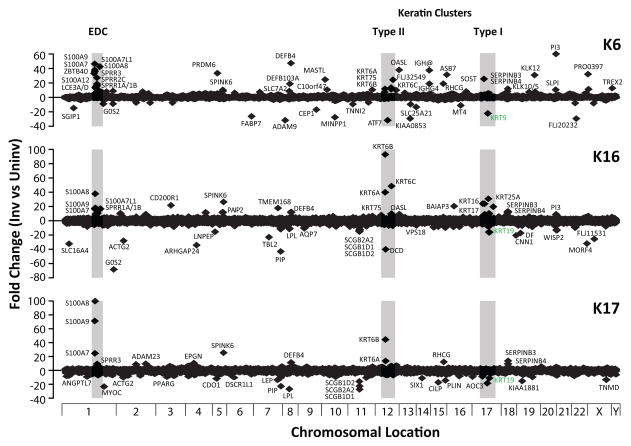
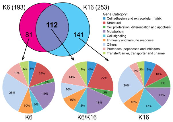
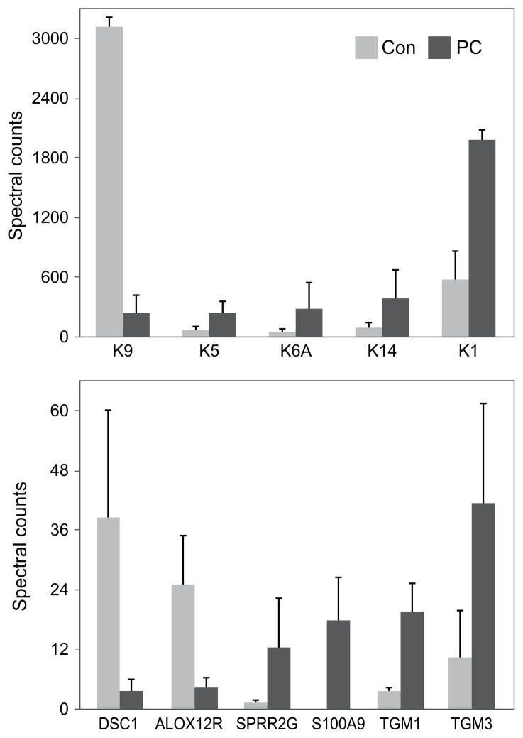
Results: A comparison of PC-involved skin biopsies to adjacent uninvolved plantar skin identified 112 differentially-expressed mRNAs common to patient groups harboring K6 (i.e., both K6a and K6b) and K16 mutations. Among these mRNAs, 25 encode structural proteins including keratins, small proline-rich and late cornified envelope proteins, 20 are related to metabolism and 16 encode proteases, peptidases, and their inhibitors including kallikrein-related peptidases (KLKs), and serine protease inhibitors (SERPINs). mRNAs were also identified to be differentially expressed only in K6 (81) or K16 (141) patient samples. Furthermore, 13 mRNAs were identified that may be involved in pain including nociception and neuropathy. Protein profiling, comparing three K6a plantar tape-stripping samples to non-PC controls, showed changes in the PC corneocytes similar, but not identical, to the mRNA analysis.

Conclusion: Many differentially-expressed genes identified in PC-involved skin encode components critical for skin barrier homeostasis including keratinocyte proliferation, differentiation, cornification, and desquamation. The profiling data provide a foundation for unraveling the pathogenesis of PC and identifying targets for developing effective PC therapeutics.

Keywords: Desquamation; Genodermatosis; Keratinocyte; Monogenic skin disorder; Painful palmoplantar keratoderma; mTOR signaling pathway.


Conflict of interest statement

Conflict of Interest

None.

Figures

Fig. 1
Fig. 1. Physical locations of plantar biopsy sites for one of the participating patients (K16-R127C mutation)
Sites (involved and uninvolved) where biopsies were obtained (as described in Methods and Materials) are circled. Similar biopsy pairs were collected from all PC participants and non-PC volunteers (controls).
Fig. 2
Fig. 2. Chromosomal location and hotspots for differentially-expressed genes in PC skin
mRNA expression profiling results, comparing PC-involved and uninvolved skin biopsies obtained from genotyped patients, were grouped by keratin mutation (K6, n=3; K16, n=3; and K17, n=1). For each of the 18,771 unique genes represented in the microarray, the average fold change of transcripts in PC-involved skin vs. adjacent uninvolved skin was plotted against chromosomal location. Genes whose expression levels are markedly altered (usually by greater than 8-fold) are labeled. Three hotspots for highly differentially-expressed genes are shaded.
Fig. 3
Fig. 3. Venn diagram and classification of differentially-expressed genes by genotype
mRNA profiling results were divided into K6 and K16 groups according to patient genotype. Genes satisfying the criteria of fold change ≥ 3.5 and p-value ≤ 0.1 (when compared to matched PC-uninvolved skin biopsies) were identified as differentially expressed (see Materials and Methods). Of the resulting 193 genes in K6 patients (n=3) and 253 genes in K16 patients (n=3), 112 are common to both K6 and K16 patient groups, while 81 genes are differentially expressed only in K6 patients and 141 genes only in K16 patients. The identified genes were classified into different categories with color coding (applies to pie chart) according to function.
Fig. 4
Fig. 4. Validation of mRNA profiling results by RT-qPCR and immunohistochemistry
Selected differentially-expressed genes identified by mRNA profiling that are potentially related to nociception and neuropathy were further analyzed by RT-qPCR to confirm the mRNA profiling findings. For each gene analyzed, RT-qPCR was performed (> three replicates) and the average fold change of expression levels (involved vs. uninvolved) was plotted as mean ± standard deviation (n=4) (A). SPRR1A was selected for further investigation by immunohistochemistry of frozen skin sections (10 μm). Increased protein expression of SPRR1a (red) is observed in PC-involved skin biopsies (B) compared to uninvolved (D). Enlarged images of areas marked by squares are also shown (C and E). Nuclei were counterstained with DAPI (blue) Scale bar = 500 μm.
Fig. 5
Fig. 5. Differences in protein profile between PC and normal epidermis
Illustrated are the normalized weighted spectral counts of keratins (top) and other prominent keratinocyte proteins (bottom) from three subjects, each harboring the K6a N171K mutation.
Fig. 6
Fig. 6. Proposed model of molecular pathways involved in PC pathogenesis
In healthy skin, keratinocyte cornification and desquamation exist in equilibrium to maintain the appropriate epithelium thickness. In PC, this process is imbalanced as exemplified by aconthosis associated with increased levels of structural (keratins, SPRRs, IVL, and late cornified envelope) and adhesion (CDSN, DSC2, and GJB2) proteins in the SC. Desquamation is putatively delayed by inhibition of key desquamation enzymes such as KLK5, KLK7, and cathepsins, likely through increased production of protease inhibitors including SPINK6, SPINK5, elafin, SLPI, and SERPINs. This imbalance likely leads to the observed keratoderma. Mechanical stress acting on the altered skin barrier may trigger pain, potentiated by mTOR pathway activation, increased KLK10 and bradykinin levels and/or tissue damage caused by microbial colonization and infection in the skin.

References

    1. Bowden PE, Haley JL, Kansky A, Rothnagel JA, Jones DO, Turner RJ. Mutation of a type II keratin gene (K6a) in pachyonychia congenita. Nat Genet. 1995;10:363–365. - PubMed
    1. McLean WH, Rugg EL, Lunny DP, Morley SM, Lane EB, Swensson O, et al. Keratin 16 and keratin 17 mutations cause pachyonychia congenita. Nat Genet. 1995;9:273–278. - PubMed
    1. Smith FJ, Jonkman MF, van Goor H, Coleman CM, Covello SP, Uitto J, et al. A mutation in human keratin K6b produces a phenocopy of the K17 disorder pachyonychia congenita type 2. Hum Mol Genet. 1998;7:1143–1148. - PubMed
    1. Leachman SA, Kaspar RL, Fleckman P, Florell SR, Smith FJ, McLean WH, et al. Clinical and pathological features of pachyonychia congenita. J Investig Dermatol Symp Proc. 2005;10:3–17. - PubMed
    1. McLean WH, Hansen CD, Eliason MJ, Smith FJ. The phenotypic and molecular genetic features of pachyonychia congenita. The Journal of investigative dermatology. 2011;131:1015–1017. - PubMed

Publication types