

Long-term survival after thoracoscopic enucleation of a gastrointestinal stromal tumor arising from the esophagus
- PMID: 25656166
- PMCID: PMC4318488
- DOI: 10.1093/jscr/rju155
Long-term survival after thoracoscopic enucleation of a gastrointestinal stromal tumor arising from the esophagus
Abstract
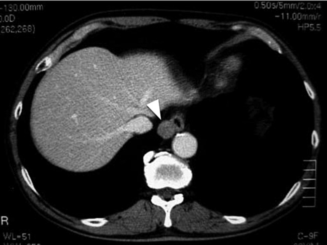
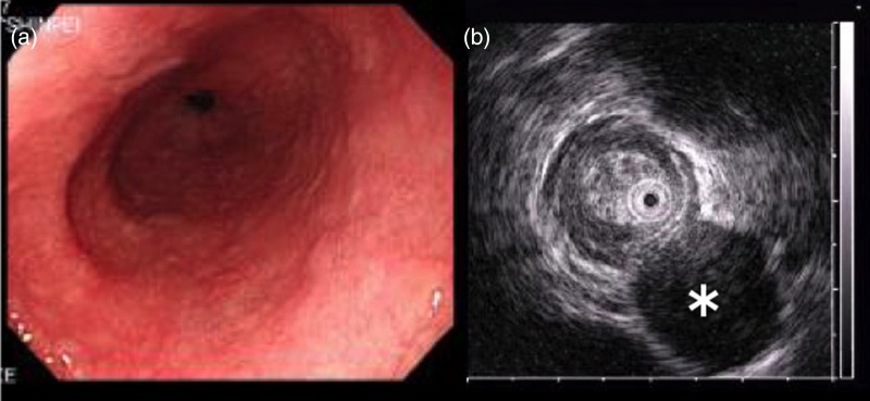
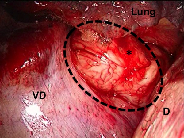
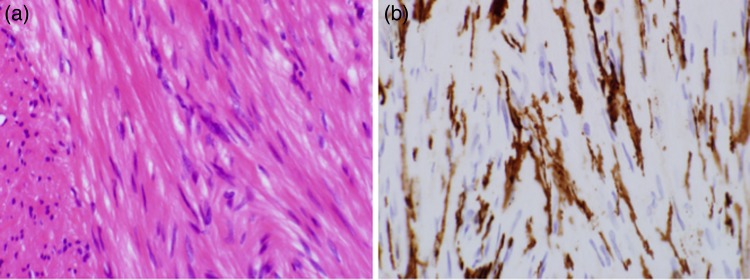
The goal of surgical treatment for gastrointestinal stromal tumor (GIST) is the complete resection of the tumor. A 62-year-old male had a clearly distinguishable mass having a smooth surface at the right side of the lower esophagus by computed tomography. Thoracoscopic resection of the tumor was performed. Immunohistochemical analysis showed that the tumor was positive for c-KIT and CD34 without mitosis, and diagnosed to be a low-risk GIST. At 6 years after surgery, the patient survived without recurrence. This study described the long-term surviving patient without the recurrence of tumor after the thoracoscopic resection of an esophageal GIST.
Published by Oxford University Press and JSCR Publishing Ltd. All rights reserved. © The Author 2015.
Figures
References
-
- Demetri GD, Benjamin RS, Blanke CD, Choi H, Corless C, DeMatteo RP, et al. NCCN task force report: optimal management of patients with gastrointestinal stromal tumor (GIST): expansion and update of NCCN clinical practice guidelines. J Natl Compr Canc Netw. 2004;Suppl 1:S1–26.. - PubMed
-
- Gupta P, Tewari M, Shukla HS. Gastrointestinal stromal tumor. Surg Oncol. 2008;17:129–38. - PubMed
-
- Koide N, Kishimoto K, Komatsu O, Yoshizawa A, Sugiyama A, Miyagawa S. Thoracoscopic enucleation of esophageal stromal tumor. Dis Esophagus. 2004;17:104–8. - PubMed
Publication types
LinkOut - more resources
Full Text Sources
Other Literature Sources
