. 2015 May;45(3):313-23.
doi: 10.1007/s10519-015-9708-8. Epub 2015 Feb 7.

Longitudinal development of hormone levels and grey matter density in 9 and 12-year-old twins

Affiliations

Longitudinal development of hormone levels and grey matter density in 9 and 12-year-old twins

Rachel M Brouwer et al. Behav Genet. 2015 May.

Abstract

Puberty is characterized by major changes in hormone levels and structural changes in the brain. To what extent these changes are associated and to what extent genes or environmental influences drive such an association is not clear. We acquired circulating levels of luteinizing hormone, follicle stimulating hormone (FSH), estradiol and testosterone and magnetic resonance images of the brain from 190 twins at age 9 [9.2 (0.11) years; 99 females/91 males]. This protocol was repeated at age 12 [12.1 (0.26) years] in 125 of these children (59 females/66 males). Using voxel-based morphometry, we tested whether circulating hormone levels are associated with grey matter density in boys and girls in a longitudinal, genetically informative design. In girls, changes in FSH level between the age of 9 and 12 positively associated with changes in grey matter density in areas covering the left hippocampus, left (pre)frontal areas, right cerebellum, and left anterior cingulate and precuneus. This association was mainly driven by environmental factors unique to the individual (i.e. the non-shared environment). In 12-year-old girls, a higher level of circulating estradiol levels was associated with lower grey matter density in frontal and parietal areas. This association was driven by environmental factors shared among the members of a twin pair. These findings show a pattern of physical and brain development going hand in hand.


Figures

Fig. 1
Fig. 1
Path diagram representing the longitudinal genetic model fitted to the data collected at age 9 and 12. For simplicity, only the diagram for twin 1 is shown. The genetic components A1 to A4 of twin 1 are connected to those of twin 2 with a correlation of 1 in monozygotic twins, and 0.5 in dizygotic twins. The common environmental components C1 to C4 of twin 1 are connected to those of twin 2 with a correlation of 1 by definition. For visualization purposes, the common and unique environmental correlations and paths are not labeled. The submodel that was used to investigate the association between estradiol and grey matter density at age 12 only is colored green (Color figure online)
Fig. 2
Fig. 2
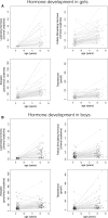
Changes in hormone levels over time in girls (a) and boys (b) (Color figure online)
Fig. 3
Fig. 3
Significant positive associations between changes in grey matter density and changes in FSH levels in girls (FDR corrected; alpha = 0.05). Clusters of positive associations were found in the left hippocampus (middle panel inset), left frontal areas (top and middle panel), left precuneus (top and bottom panel), and right cerebellum (bottom panel). Bottom right the average correlation between changes in grey matter density and changes in FSH split up into the contribution of genetic influences shared by the two phenotypes (rph-a), common environmental influences shared by the two phenotypes (rph-c) and contribution of unique environmental influences shared by the two phenotypes (rph-e). These three add up to the observed phenotypic correlation (Color figure online)
Fig. 4
Fig. 4
Significant negative associations between grey matter density and estradiol levels in girls at age 12 (FDR corrected; alpha = 0.05). Associations were predominantly found in (left) frontal and parietal areas (Color figure online)

References

    1. Bay K, Andersson AM, Skakkebaek NE. Estradiol levels in prepubertal boys and girls—analytical challenges. Int J Androl. 2004;27(5):266–273. doi: 10.1111/j.1365-2605.2004.00487.x. - DOI - PubMed
    1. Boker S, Neale M, Maes H, Wilde M, Spiegel M, Brick T, Spies J, Estabrook R, Kenny S, Bates T, Mehta P, Fox J. OpenMx: an open source extended structural equation modeling framework. Psychometrika. 2011;76:306–317. doi: 10.1007/s11336-010-9200-6. - DOI - PMC - PubMed
    1. Boomsma DI, Busjahn A, Peltonen L. Classical twin studies and beyond. Nat Rev Genet. 2002;3:872–882. doi: 10.1038/nrg932. - DOI - PubMed
    1. Bramen JE, Hranilovich JA, Dahl RE, Forbes EE, Chen J, Toga AW, Dinov ID, Worthman CM, Sowell ER. Puberty influences medial temporal lobe and cortical grey matter maturation differently in boys than girls matched for sexual maturity. Cereb Cortex. 2011;21:636–646. doi: 10.1093/cercor/bhq137. - DOI - PMC - PubMed
    1. Bramen JE, Hranilovich JA, Dahl RE, Chen J, Rosso C, et al. Sex matters during adolescence: testosterone-related cortical thickness maturation differs between boys and girls. PLoS ONE. 2012;7(3):e33850. doi: 10.1371/journal.pone.0033850. - DOI - PMC - PubMed

Publication types

LinkOut - more resources