. 2015 Apr;19(4):826-35.
doi: 10.1111/jcmm.12510. Epub 2015 Feb 5.

Rosiglitazone via PPARγ-dependent suppression of oxidative stress attenuates endothelial dysfunction in rats fed homocysteine thiolactone

Affiliations

Rosiglitazone via PPARγ-dependent suppression of oxidative stress attenuates endothelial dysfunction in rats fed homocysteine thiolactone

Xu-Hong Yang et al. J Cell Mol Med. 2015 Apr.

Abstract

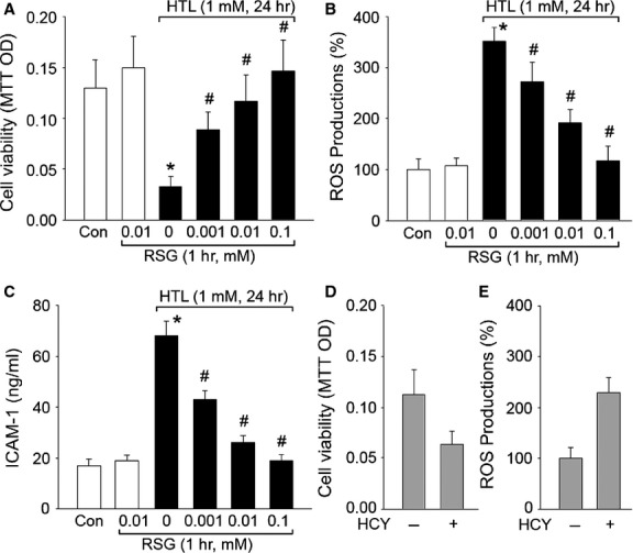
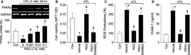
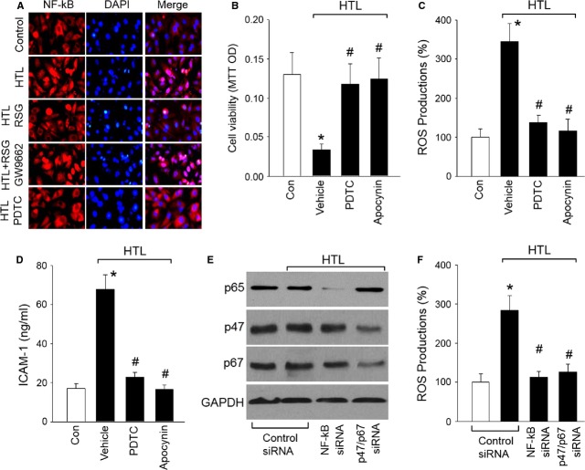
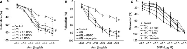
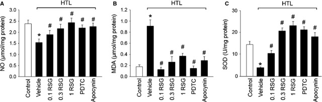
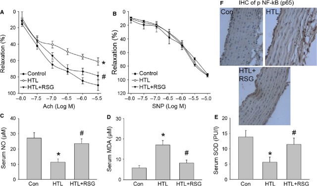
To explore whether rosiglitazone (RSG), a selective peroxisome proliferator-activated receptor γ (PPARγ) agonist, exerts beneficial effects on endothelial dysfunction induced by homocysteine thiolactone (HTL) and to investigate the potential mechanisms. Incubation of cultured human umbilical vein endothelial cells with HTL (1 mM) for 24 hrs significantly reduced cell viabilities assayed by 3-(4,5-dimethyl-2-thiazolyl)-2,5-diphenyl-2-H-tetrazolium bromide, as well as enhanced productions of reactive oxygen species, activation of nuclear factor kappa B, and increased intercellular cell adhesion molecule-1 secretion. Pre-treatment of cells with RSG (0.001-0.1 mM), pyrollidine dithiocarbamate (PDTC, 0.1 mM) or apocynin (0.1 mM) for 1 hr reversed these effects induced by HTL. Furthermore, co-incubation with GW9662 (0.01 mM) abolished the protective effects of RSG on HTL-treated cells. In ex vivo experiments, exposure of isolated aortic rings from. rats to HTL (1 mM) for 1 hr dramatically impaired acetylcholine-induced endothelium-dependent relaxation, reduced release of nitric oxide and activity of superoxide dismutase, and increased malondialdehyde content in aortic tissues. Preincubation of aortic rings with RSG (0.1, 0.3, 1 mM), PDTC or apocynin normalized the disorders induced by HTL. In vivo analysis indicated that administration of RSG (20 mg/kg/d) remarkably suppressed oxidative stress and prevented endothelial dysfunction in rats fed HTL (50 mg/kg/d) for 8 weeks. RSG improves endothelial functions in rats fed HTL, which is related to PPARγ-dependent suppression of oxidative stress.

Keywords: endothelial dysfunction; homocysteine thiolactone; oxidative stress; rosiglitazone; vascular ageing.


Figures

Fig 1
Fig 1
Rosiglitazone dose-dependently suppresses HTL-induced oxidative stress and improves cell viabilities in cultured HUVECs. Cultured HUVECs were pre-treated with rosiglitazone (RSG, 0.001–1 mM) for 1 hr and then incubated with homocysteine thiolactone (HTL, 1 mM) for 24 hrs. (A) Cell viability was measured by MTT. (B) Intracellular ROS productions were detected by DHE/HPLC. (C) The concentration of soluble intercellular adhesion molecule 1 (sICAM-1) in culture medium was assayed by ELISA. All data were expressed as mean ± SEM. N is 3 in each group. *P < 0.05 versus Con, #P < 0.05 versus HTL alone. (D and E) Cultured HUVECs were treated with homocysteine (HCY, 1 mM) for 24 hrs. (D) Cell viability and (E) intracellular ROS productions were measured by MTT and DHE/HPLC respectively. Data were expressed as mean ± SEM. N is 3 in each group. *P < 0.05 versus Control.
Fig 2
Fig 2
Inhibition of PPARγ abolished rosiglitazone-suppressed oxidative stress induced by HTL in endothelial cells. (A) Cultured HUVECs were pre-treated with rosiglitazone (RSG, 0.001–1 mM) for 1 hr and then incubated with homocysteine thiolactone (HTL, 1 mM) for 24 hrs. The expression of PPARγ mRNA was examined by RT-PCR. All data were expressed as mean ± SEM. The picture is a representative picture from three independent experiments. *P < 0.05 versus Con, #P < 0.05 versus HTL alone. (B–D) Cultured HUVECs were pre-treated with rosiglitazone (RSG, 1 mM) for 1 hr and then incubated with homocysteine thiolactone (HTL, 1 mM) for 24 hrs in presence of GW9662 (0.01 mM). The expression of PPARγ mRNA was examined by RT-PCR. Cell was subjected to assay (B) Cell viability by MTT, (C) Intracellular ROS productions by DHE fluorescence and (D) The concentration of sICAM-1 in culture medium by ELISA. All data were expressed as mean ± SEM. N is 3 in each group. *P < 0.05 versus Con, &P < 0.05 versus HTL alone, $P < 0.05 versus HTL+RSG.
Fig 3
Fig 3
Rosiglitazone via PPARγ inhibits NF-κB to suppress oxidative stress and protects cell functions in endothelial cells. (A) Cultured HUVECs were pre-treated with 1 mM RSG, 1 mM RSG plus 0.01 mM GW9662 or 0.1 mM PDTC for 1 hr and then incubated with homocysteine thiolactone (HTL, 1 mM) for 24 hrs. IHC fluorescence was used to detect NF-κB activity by staining cells with primary anti-NF-κB antibody. Red, NF-κB; Blue, DAPI (nucleus). The picture is a representative picture from three independent experiments. (B–D) Cultured HUVECs were pre-treated with 0.1 mM PDTC or apocynin (0.1 mM) for 1 hr and then incubated with homocysteine thiolactone (HTL, 1 mM) for 24 hrs. Cell was subjected to assay (B) Cell viability by MTT, (C) Intracellular ROS productions by DHE fluorescence, and (D) The concentration of sICAM-1 in culture medium by ELISA. All data were expressed as mean ± SEM. N is 3 in each group. *P < 0.05 versus Con, #P < 0.05 versus HTL alone. (E and F) Cultured HUVECs were transfected with control siRNA, NF-κB (p65) siRNA and NAD(P)H oxidase (p47/p67) siRNA for 48 hrs. Then cells were treated with HTL (1 mM) for 24 hrs. (E) Protein levels of NF-κB p65, and NAD(P)H oxidase (p47 and p67) in total cell lysates and (F) intracellular ROS productions were measured by Western blot and DHE/HPLC respectively. Data were expressed as mean ± SEM. N is 3 in each group. *P < 0.05 versus Control siRNA alone. #P < 0.05 versus Control siRNA plus HTL.
Fig 4
Fig 4
Rosiglitazone attenuates HTL-impaired endothelium-dependent relaxation in rat aortas. (A) The isolated rat aortic rings were exposed to HTL (1 mM) for 1 hr after preincubation with rosiglitazone (RSG, 0.1–1 mM) for 30 min. The endothelium-dependent relaxation induced by acetylcholine (Ach) was assayed by organ chamber. (B) The isolated rat aortic rings were exposed to HTL (1 mM) for 1 hr after preincubation with 0.1 mM PDTC or apocynin (0.1 mM) for 30 min. The endothelium-dependent relaxation induced by acetylcholine (Ach) was assayed by organ chamber. All data were expressed as mean ± SEM. N is 5 in each group. *P < 0.05 versus Con, #P < 0.05 versus HTL alone. (C) Endothelium-independent relaxation was assayed in A and B by using sodium nitroprusside (SNP).
Fig 5
Fig 5
HTL via PPARγ/NF-κB/NADPH oxidase induces oxidative stress in isolated rat aortas. The isolated rat aortic rings were exposed to HTL (1 mM) for 1 hr after preincubation with rosiglitazone (RSG, 0.1–1 mM), PDTC (0.1 mM) or apocynin (0.1 mM) for 30 min. Homogenates of aortic tissues were subjected to assay (A) nitric oxide productions by Griess method, (B) MDA content by TBA method and (C) SOD activity by the spectrophotometric method. All data were expressed as mean ± SEM. N is 5 in each group. *P < 0.05 versus Con, #P < 0.05 versus HTL alone.
Fig 6
Fig 6
Rosiglitazone suppresses oxidative stress and improves endothelial functions in rats fed HTL in vivo. The rats were intragastric gavaged HTL (50 mg/kg/d) and received administration of rosiglitazone (RSG, 20 mg/kg/d) for 8 weeks. At the end of experiments, rats were killed under anaesthesia. Artery from descending aorta was subjected to assay (A) endothelium-dependent relaxation induced by acetylcholine (Ach) and (B) endothelium-independent relaxation induced by sodium nitroprusside (SNP) in organ chamber. Blood was collected to assay (C) serum nitric oxide level by Griess method, (D) serum MDA content by TBA method and (E) SOD activity by the spectrophotometric method. All data were expressed as mean ± SEM. Five to ten rats in each group. *P < 0.05 versus Con, #P < 0.05 versus HTL alone. (F) Immunohistochemical analysis of active NF-κB in arteries by using phospho-p65 antibody. The picture (×40) is a representative picture from five independent experiments.

Similar articles

Cited by

References

    1. McCully KS. Vascular pathology of homocysteinemia: implications for the pathogenesis of arteriosclerosis. Am J Pathol. 1969;56:111–28. - PMC - PubMed
    1. Kolodziejczyk-Czepas J, Talar B, Nowak P, et al. Homocysteine and its thiolactone impair plasmin activity induced by urokinase or streptokinase in vitro. Int J Biol Macromol. 2012;50:754–8. - PubMed
    1. Liu Y, Tian T, Zhang H, et al. The effect of homocysteine-lowering therapy with folic acid on flow-mediated vasodilation in patients with coronary artery disease: a meta-analysis of randomized controlled trials. Atherosclerosis. 2014;235:31–5. - PubMed
    1. de Andrade CR, Fukada SY, Olivon VC, et al. Alpha1d-adrenoceptor-induced relaxation on rat carotid artery is impaired during the endothelial dysfunction evoked in the early stages of hyperhomocysteinemia. Eur J Pharmacol. 2006;543:83–91. - PubMed
    1. Kersten S, Desvergne B, Wahli W. Roles of ppars in health and disease. Nature. 2000;405:421–4. - PubMed

Publication types

MeSH terms

LinkOut - more resources