Controlled Clinical Trial
. 2015 Sep-Oct;25(5):799-806.
doi: 10.1111/jon.12193. Epub 2015 Feb 6.

Heterogeneity of Multiple Sclerosis White Matter Lesions Detected With T2*-Weighted Imaging at 7.0 Tesla

Affiliations
Controlled Clinical Trial

Heterogeneity of Multiple Sclerosis White Matter Lesions Detected With T2*-Weighted Imaging at 7.0 Tesla

Bing Yao et al. J Neuroimaging. 2015 Sep-Oct.

Abstract

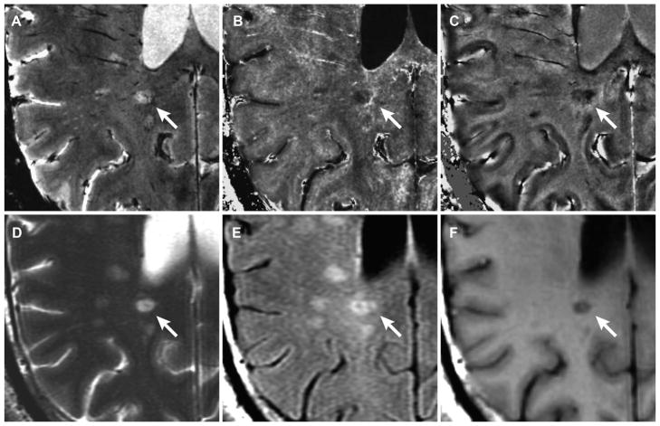
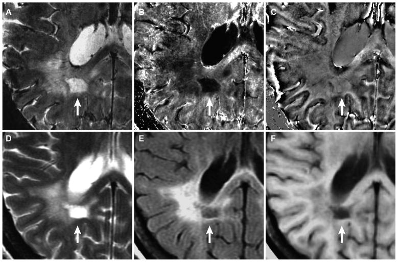
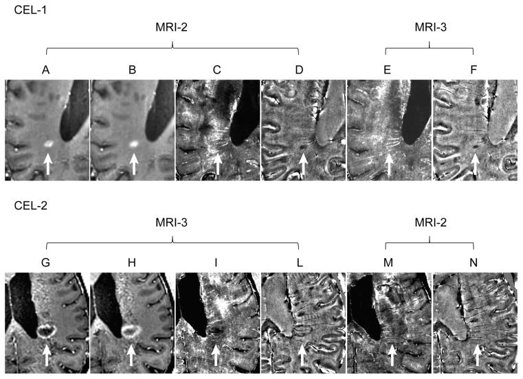
Background and purpose: Postmortem studies in multiple sclerosis (MS) indicate that in some white matter lesions (WM-Ls), iron is detectable with T2*-weighted (T2*-w), and its reciprocal R2* relaxation rate, magnetic resonance imaging (MRI) at 7.0 Tesla (7T). This iron appears as a hyperintense rim in R2* images surrounding a hypointense core. We describe how this observation relates to clinical/radiological characteristics of patients, in vivo.

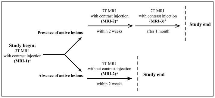
Methods: We imaged 16 MS patients using 3T and 7T scanners. WM-Ls were identified on T1-w / T2-w 3T-MRIs. Thereafter, WM-Ls with a rim of elevated R2* at 7T were counted and compared to their appearance on conventional MRIs.

Results: We counted 36 WM-Ls presenting a rim of elevated R2* in 10 patients. Twenty-three (64%) lesions coincided with focal WM-Ls on T2-w MRIs; 13 (36%) coincided with only portions of larger lesions on T2-w images; and 20 (56%) corresponded to a hypointense chronic black hole. WM-Ls presenting a rim of elevated R2* were seen in both relapsing-remitting patients with low disability and in those with long-standing secondary progressive MS.

Conclusions: WM-Ls with a contour of high R2* are present at different MS stages, potentially representing differences in the contribution of iron in MS disease evolution.

Keywords: 7.0 tesla; Multiple sclerosis; T2*-weighted; iron; magnetic resonance imaging.


Conflict of interest statement

Conflicts of Interest: None of the authors has any conflicts with the work.

Figures

Fig. 1
Fig. 1
Study design. Each person underwent a 3T (MRI-1) and a 7T MRI (MRI-2) scan obtained 2 weeks apart. In patients only, when a CEL was noted in postcontrast imaging of MRI-1, postcontrast sequences were obtained at the MRI-2 7T scan. The 7T scan, inclusive of pre-and postcontrast sequences, was also repeated a month later (MRI-3). On the same day of either MRI-1 or MRI-2, each patient underwent a clinical assessment to establish the EDSS score. * Indicates the time when the clinical assessment was performed. See text for detailed explanation.
Fig. 2
Fig. 2
WM-L with a rim of increased signal in R2*. WM-L with a rim of increased signal in R2* (white arrow) identified in magnitude (A), R2* (B), and phase (C) map in a RRMS patient with 1.5 years of MS and EDSS score of 2.5 (patient-2 in Tables 2 and 3). The lesion entirely coincides with a focal lesion in T2-w FSE (D), T2-w FLAIR (E) image, and T1-cBH in T1-w SE image (F) at 3T.
Fig. 3
Fig. 3
WM-L with a rim of increased signal in R2* WM-L with a rim of increased signal in R2* (white arrow) identified in magnitude (A), R2* (B), and phase (C) map in a SPMS patient with 33 years of disease and EDSS score of 6.0 (patient-5 in Tables 2 and 3). The lesion is only part of a larger lesion seen in T2-w FSE (D), T2-w FLAIR (E) image, and T1-w SE (F) image. The lesion also corresponds with a black hole on FLAIR image, indicating end-stage cavitation.
Fig. 4
Fig. 4
Active lesions with and without a rim of elevated R2 CEL-1 (white arrow) seen in the T1-w MPRAGE of MRI-1 obtained 5 minutes post-Gd injection (A) and 40 minutes post-Gd injection (B). The R2* and phase maps show the central core of the enhancing lesion had an decreased R2* (C) and a reduced phase (ie, paramagnetic shift) (D). No clear signal alterations ascribable to the prior presence of that CEL could be unambiguously identified in the R2* map of MRI-3 (E) obtained a month later when the CEL was no longer enhancing. Residual smaller area with a paramagnetic phase shift was visible in the phase map (F) of MRI-2. CEL-2 in MRI-3 is indicated with white arrow. This CEL presented with a ring-enhancing pattern in the T1-w MPRAGE obtained 5 minutes (G) 40 minutes post-Gd injection (H). The ring-enhancing contour coincided with a rim of elevated R2* signal (I) and paramagnetic phase shift (L). Minimal signs of signal alterations corresponding to the presence of this lesion were present in the R2* (M) but not on the phase (N) images obtained a month earlier. The fainting nature of this signal may signify an artifact. One cannot, however, unambiguously exclude minimal brain barrier leakage and iron deposition associated with it. These data were derived from a patient with RRMS, 7.5 years of disease and EDSS score of 2.5 (patient-8 in Tables 2 and 3).

References

    1. Lassmann H. The pathologic substrate of magnetic resonance alterations in multiple sclerosis. Neuroimag Clin N Am. 2008;18:563–576. - PubMed
    1. Yao B, Li TQ, Gelderen P, et al. Susceptibility contrast in high field MRI of human brain as a function of tissue iron content. NeuroImage. 2009;44:1259–1266. - PMC - PubMed
    1. Shmueli K, de Zwart JA, van Gelderen P, et al. Magnetic susceptibility mapping of brain tissue in vivo using MRI phase data. Magn Reson Med. 2009;62:1510–1522. - PMC - PubMed
    1. Fukunaga M, Li TQ, van Gelderen P, et al. Layer-specific variation of iron content in cerebral cortex as a source of MRI contrast. Proc Natl Acad Sci USA. 2010;107:3834–3839. - PMC - PubMed
    1. Bagnato F, Hametner S, Yao B, et al. Tracking iron in multiple sclerosis: a combined imaging and histopathological study a 7 tesla. Brain. 2011;134:3602–3615. - PMC - PubMed

Publication types

LinkOut - more resources