

Genital Ulcerative Pyoderma Gangrenosum in Behçet's Disease: A Case Report and Review of the Literature
- PMID: 25657430
- PMCID: PMC4318036
- DOI: 10.4103/0019-5154.147866
Genital Ulcerative Pyoderma Gangrenosum in Behçet's Disease: A Case Report and Review of the Literature
Abstract
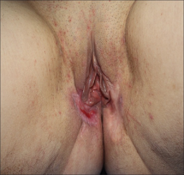
Behçet's disease (BD), first described by Hulusi Behcet, is a multisystemic disease characterized by recurrent oral and genital ulcerations, ocular and cutaneous lesions, arthritis and vascular disease. Pyoderma gangrenosum (PG) is a rare, chronic, sterile pustular and progressive ulcerative process of unknown cause; sometimes can participate in the differential diagnosis of Behcet's ulceration. A 33-year-old woman complained a severe genital ulcer. She had a purulent oozing and stinky ulceration on the right side of labium minor measuring 5-8 cm. A punch biopsy at ulcer margin showed that the lymphocytic panniculitis was extending to the subcutaneous fat tissue without fibrin deposition or necrotic changes in the vessel wall. Based on the clinical and histological findings, she was diagnosed as genital ulcerative PG, which occurred during the exacerbation of BD.
Keywords: Behçet's disease; genital ulcer; ulcerative pyoderma gangrenosum.
Conflict of interest statement
Figures


Similar articles
-
Intestinal Behcet's disease with pyoderma gangrenosum: a case report.World J Gastroenterol. 2006 Feb 14;12(6):979-81. doi: 10.3748/wjg.v12.i6.979. World J Gastroenterol. 2006. PMID: 16521233 Free PMC article.
-
Case of pyoderma gangrenosum showing oral and genital ulcers, misdiagnosed as Behcet's disease at first medical examination.J Dermatol. 2008 May;35(5):289-92. doi: 10.1111/j.1346-8138.2008.00468.x. J Dermatol. 2008. PMID: 18477229
-
Exacerbation of Behcet's Disease and Pyoderma Gangrenosum Following COVID-19 Infection: A Case Report.Cureus. 2023 Nov 25;15(11):e49386. doi: 10.7759/cureus.49386. eCollection 2023 Nov. Cureus. 2023. PMID: 38146565 Free PMC article.
-
Behçet's disease-like presentation of bullous pyoderma gangrenosum associated with Crohn's disease.Clin Exp Dermatol. 2006 May;31(3):384-6. doi: 10.1111/j.1365-2230.2006.02093.x. Clin Exp Dermatol. 2006. PMID: 16681583 Review.
-
Noninfectious genital ulcers.Semin Cutan Med Surg. 2015 Dec;34(4):187-91. doi: 10.12788/j.sder.2015.0168. Semin Cutan Med Surg. 2015. PMID: 26650697 Review.
Cited by
-
Rare pyoderma gangrenosum correlated with systemic lupus erythematosus: A case report.Clin Case Rep. 2023 Nov 2;11(11):e7857. doi: 10.1002/ccr3.7857. eCollection 2023 Nov. Clin Case Rep. 2023. PMID: 37927989 Free PMC article.
-
Pathergy phenomenon: an important clinical pointer to Behҫet disease.BMJ Case Rep. 2019 Jun 24;12(6):e230662. doi: 10.1136/bcr-2019-230662. BMJ Case Rep. 2019. PMID: 31239320 Free PMC article. No abstract available.
-
Pyoderma gangrenosum associated with Behçet's disease.Arch Rheumatol. 2020 Dec 10;36(1):140-141. doi: 10.46497/ArchRheumatol.2021.8010. eCollection 2021 Mar. Arch Rheumatol. 2020. PMID: 34046581 Free PMC article. No abstract available.
-
Scrotal Pyoderma Gangrenosum Associated with Evans Syndrome.J Clin Med. 2018 Aug 22;7(9):230. doi: 10.3390/jcm7090230. J Clin Med. 2018. PMID: 30131462 Free PMC article.
-
Microarray analysis of potential genes in the pathogenesis of recurrent oral ulcer.Int J Clin Exp Pathol. 2015 Oct 1;8(10):12419-27. eCollection 2015. Int J Clin Exp Pathol. 2015. PMID: 26722428 Free PMC article.
References
-
- International Study Group for Behçet's disease. Criteria for diagnosis of Behçet's disease. International study group for Behçet's disease. Lancet. 1990;335:1078–80. - PubMed
-
- Wolff K, Stingl G. Pyoderma gangrenosum. In: Freedberg IM, Eisen AZ, Wolff K, Austen KF, Goldsmith LA, Katz SI, editors. Fizpatrick's Dermatology in General Medicine. 6th ed. Vol. 1. New York: McGraw-Hill; 2003. pp. 969–76.
-
- Powell FC, Su WP, Perry HO. Pyoderma gangrenosum: Classification and management. J Am Acad Dermatol. 1996;34:395–409. - PubMed
-
- Kaklamani VG, Vaiopoulos G, Kaklamanis PG. Behçet's disease. Semin Arthritis Rheum. 1998;27:197–21. - PubMed
-
- Munro CS, Cox NH. Pyoderma gangrenosum associated with Behçet's syndrome - Response to thalidomide. Clin Exp Dermatol. 1988;13:408–10. - PubMed
LinkOut - more resources
Full Text Sources
Other Literature Sources