. 2015 Jan 22:9:1.
doi: 10.3389/fnana.2015.00001. eCollection 2015.

The spino-bulbar-cerebellar pathway: organization and neurochemical properties of spinal cells that project to the lateral reticular nucleus in the rat

Affiliations

The spino-bulbar-cerebellar pathway: organization and neurochemical properties of spinal cells that project to the lateral reticular nucleus in the rat

Zilli Huma et al. Front Neuroanat. .

Abstract

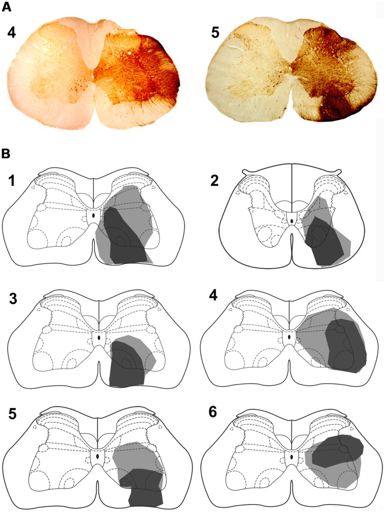
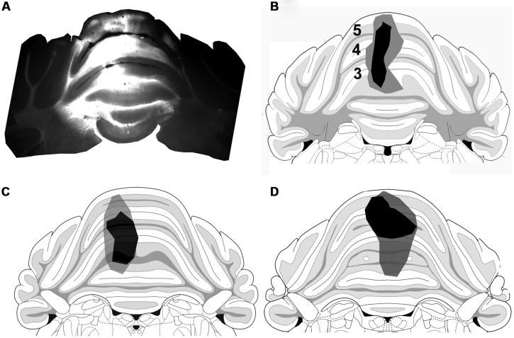
In addition to classical spinocerebellar pathways, the cerebellum receives information from the spinal cord indirectly via spino-bulbar-cerebellar systems. One of the structures in this pathway is the lateral reticular nucleus (LRt). We performed series of experiments to investigate the organization and neurotransmitter content of spinoreticular tract (SRT) neurons in the lumbar spinal cord that project to the LRt. Three rats received injections of the b subunit of Cholera toxin (CTb) or Fluorogold (FG) within the left and right LRt. The majority of SRT cells (56-61%) were found within the contralateral medial intermediate gray matter where small numbers (7-10%) of double-labeled cells were also present on both sides of the cord. Six rats received unilateral spinal injections of CTb to label spinal projections to the LRt. Injections of FG were made also into the anterior lobe of the cerebellum to label LRt pre-cerebellar neurons. Terminals were found mainly ipsilateral to spinal injection sites within the central and ventrolateral regions of the LRt. Immunocytochemical analysis of SRT terminals revealed that the majority (75%) were contained vesicular glutamate transporter 2 but a minority (20%) contained the vesicular GABA transporter. The inhibitory subpopulation was found to be GABAergic, glycinergic, or contained both transmitters. Inhibitory and excitatory terminals were present within overlapping regions of the nucleus. Most CTb terminals contacting LRt pre-cerebellar neurons were excitatory (80%) whereas a minority were inhibitory and most cells (88%) received contacts from both inhibitory and excitatory terminals. This study shows that SRT axons in the LRt have the capacity to exert direct excitatory and inhibitory actions on LRt pre-cerebellar neurons. Thus spinal cord input has the capacity to facilitate or depress the activity of individual LRt cells which in turn adjust activity in the cerebellum to produce coordinated motor behaviors.

Keywords: cerebellum; motor control; neuroanatomy; neurotransmitters; spinal cord.


Figures

FIGURE 1
FIGURE 1
Injections sites in the medulla. (A) A CTb injection site (B) An adjacent section showing a combined dark-field and epifluorescence image showing Fluorogold (FG) within the LRt and surrounding reticular formation. A schematic version of these injections is shown in Figure 2A. LRt, lateral reticular nucleus, mlf, medial longitudinal fasciculus, py, pyramid.
FIGURE 2
FIGURE 2
Bilateral medullary injection sites and distribution of spinoreticular tract (SRT) cells in lumbar segments from 3 animals (A–C). FG injections are shown as yellow and CTb as black. Diffuse spread of tracer is shown as gray. Distribution of cells for three segments (L3–L5) is shown on schematic diagrams of transverse sections. Each diagram represents a composite analysis of 3 μm × 50 μm sections from the three segments. Green stars, FG labeled cells; Red circles, CTb labeled cells; black dots, cells labeled with both tracers.
FIGURE 3
FIGURE 3
Confocal microscope images of spinal cells labeled with FG (green) and CTb (red). The main plate is a tiled image of an entire L3 transverse section. The areas demarcated by boxes 1 and 2 illustrate examples of double labeled cells within laminae VII and V, respectively, and are shown as single optical sections at higher magnification below (1 and 2). Scale bars: Main plate = 200 μm; 1 and 2 = 20 μm.
FIGURE 4
FIGURE 4
Spinal injection sites. (A) Photomicrograps of the L4 spinal segment showing CTb injection site for animals 4 and 5. The drawings (B) show the spread of CTb within the gray and white matter. The dark region represents the core of the injection and the gray region the spread of CTb. Spinal injections are in L4 with the exception of animal 2 which was in T11. Note that the core of most injections is within the ventral quadrant of the cord except for animal 6 where it was in the intermediate gray matter. Animals 1–3 were also used in experiments to label precerebellar cells.
FIGURE 5
FIGURE 5
(A) A photomicrograph showing a coronal section through the medulla the areas demarcated by boxes on the left and the right are shown at high magnification in (B) and (C), respectively, which show terminal labeling within the LRt contralateral and ipsilateral to the spinal injection site. Note the particularly large numbers of terminals in the ipsilateral LRt. (D) A representation of A (based on Paxinos and Watson, 2005) with terminal locations plotted to show their relationships with medullary structures (right = contralateral to spinal injection; -14.4 relative to Bregma). LRt, lateral reticular nucleus; LRtPC lateral reticular nucleus, parvicellular part; MdV, medullary reticular nucleus; mlf, medial longitudinal fasciculus; py, pyramid. Scale bar for (B,C) = 100 μm.
FIGURE 6
FIGURE 6
Single optical sections showing immunochemical properties of axon terminals projecting from the lumbar spinal cord in the lateral reticular nucleus ipsilateral to the spinal injection. (A) An overview of terminals within the LRt labeled for CTb, VGLUT1+2, and VGAT. The area demarcated by the box is shown in series (B,C). (B) CTb terminals (red) that are labeled for VGLUT1+2 (B’,B”). (C) CTb terminals that are labeled for VGAT (C’,C”). A merged image from both series is shown on the right adjacent to (B”). (D) CTb terminals that are labeled for GlyT2 (D’) and GAD (D”). (E) Terminals that are labeled for GlyT2 (E’) but not GAD (E”). (F) Terminals that are not labeled for GlyT2 (F’) but are labeled for GAD (F”). Arrows indicate double-labeled structures in each series. VGLUT1+2, vesicular glutamate transporter 1 and vesicular glutamate transporter 2; VGAT, vesicular GABA transporter; GLYT2, glycine transporter 2; GAD glutamate decarboxylase. Scale bars (A) = 10 μm, (B,C) = 5 μm, (D–F) = 5 μm.
FIGURE 7
FIGURE 7
Cerebellum injection sites. (A) A fluorescence micrograph showing a FG injection site within a coronal section of the anterior cerebellum. (B–D) Drawings of the core (black) and spread (gray) of FG within lobules 3, 4, and 5 of animals 1–3, respectively, (based on Paxinos and Watson, 2005).
FIGURE 8
FIGURE 8
Immunochemical properties of contacts on pre-cerebellar neurons in the LRt. (A) A fluorescent micrograph showing a coronal section through the medulla containing pre-cerebellar neurons labeled with FG from the anterior cerebellum. Note the cells within the LRt and the inferior olivary complex (IO). (Gr, Gracile nucleus; mlf, medial longitudinal fasciculus; py, pyramid; Sp5, spinal trigeminal nucleus). (B) A single optical section showing a precerebellar neuron (green) with several CTb (red) contacts on it. The contact shown in the box (C) is immunoreactive for VGAT (C–C”) and the contact shown in box (D) is immunoreactive for VGLUT2 (D–D”). CTb, b subunit of cholera toxin; FG, Fluorogold; VGLUT2, vesicular glutamate transporter 2; VGAT, vesicular GABA transporter. Scale Bars (B) = 10 μm; (C,D) = 5 μm.

References

    1. Alstermark B., Ekerot C. F. (2013). The lateral reticular nucleus: a precerebellar centre providing the cerebellum with overview and integration of motor functions at systems level. A new hypothesis. J. Physiol. 591 5453–5458 10.11/13/jphysiol.2013.256669 - DOI - PMC - PubMed
    1. Araki T., Yamano M., Murakami T., Wanaka A., Betz H., Tohyama M. (1988). Localization of glycine receptors in the rat central nervous system: an immunocytochemical analysis using monoclonal antibody. Neuroscience 25 613–624 10.1016/0306-4522(88)90263-1 - DOI - PubMed
    1. Arshavsky Y., Gelfand I. M., Orlovsky G. N., Pavlova G. A., Popova L. B. (1984). Origin of signals conveyed by the ventral spino-cerebellar tract and spino-reticulo-cerebellar pathway. Exp. Brain Res. 54 426–431 10.1007/BF00235467 - DOI - PubMed
    1. Brodal A. (1949). Spinal afferents to the lateral reticular nucleus of the medulla oblongata in the cat; an experimental study. J. Comp. Neurol. 91 259–295 10.1002/cne.900910206 - DOI - PubMed
    1. Chen S., Aston-Jones G. (1995). Evidence that cholera toxin B subunit (CTb) can be avidly taken up and transported by fibers of passage. Brain Res. 674 107–111 10.1016/0006-8993(95)00020-Q - DOI - PubMed

LinkOut - more resources