Review
. 2015 Jan 22:5:688.
doi: 10.3389/fimmu.2014.00688. eCollection 2014.

Sensing of Pyrophosphate Metabolites by Vγ9Vδ2 T Cells

Affiliations
Review

Sensing of Pyrophosphate Metabolites by Vγ9Vδ2 T Cells

Siyi Gu et al. Front Immunol. .

Abstract

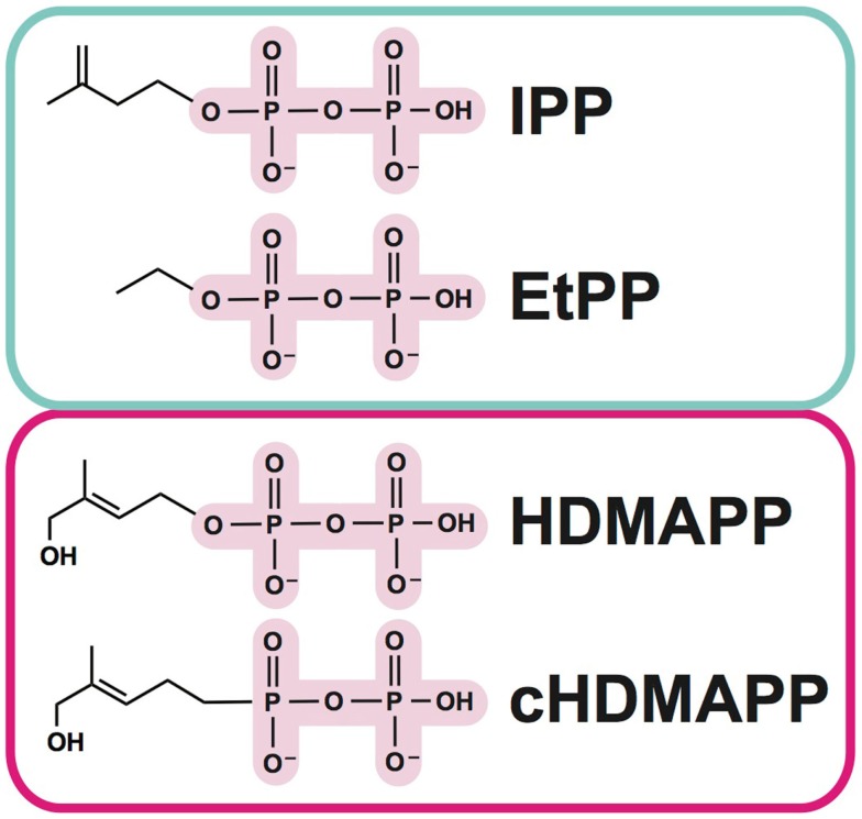
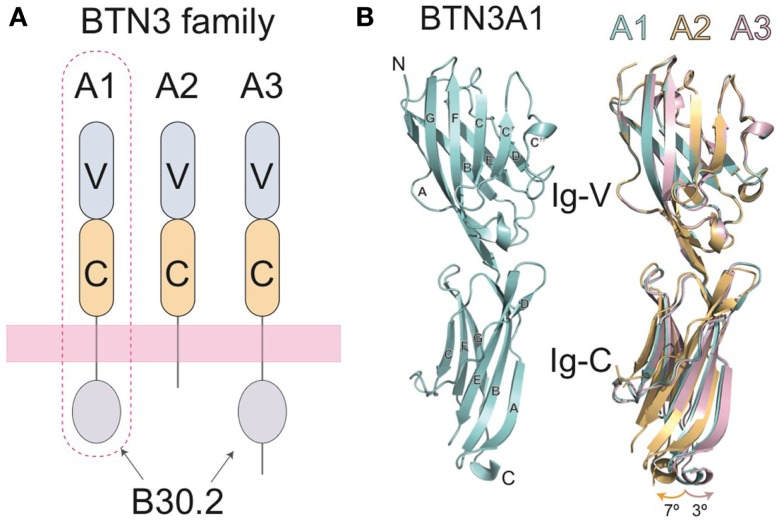
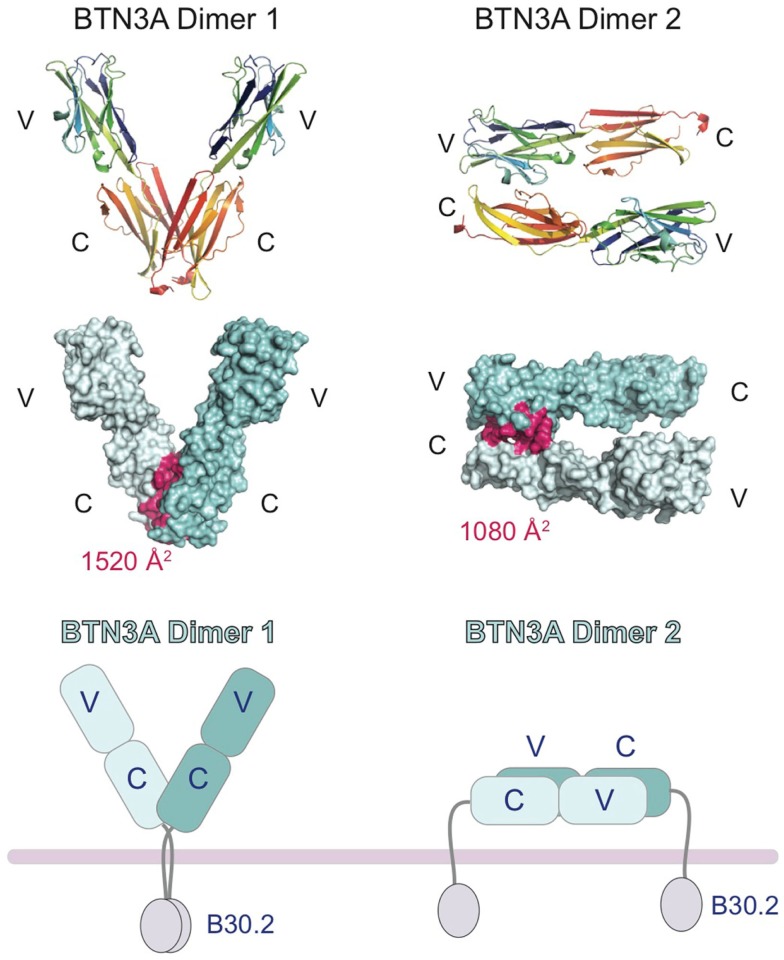
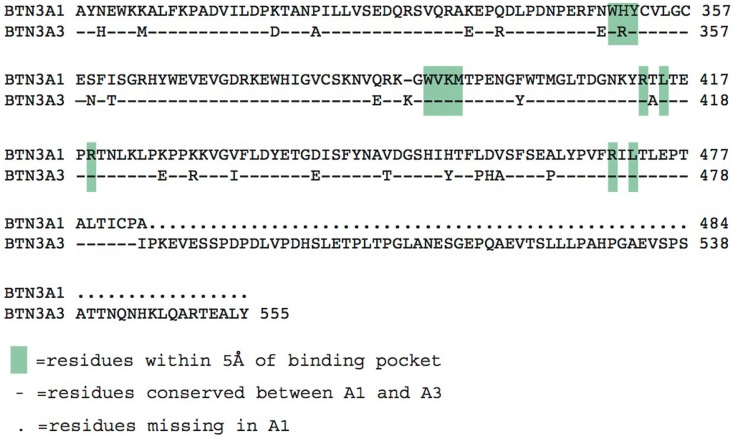
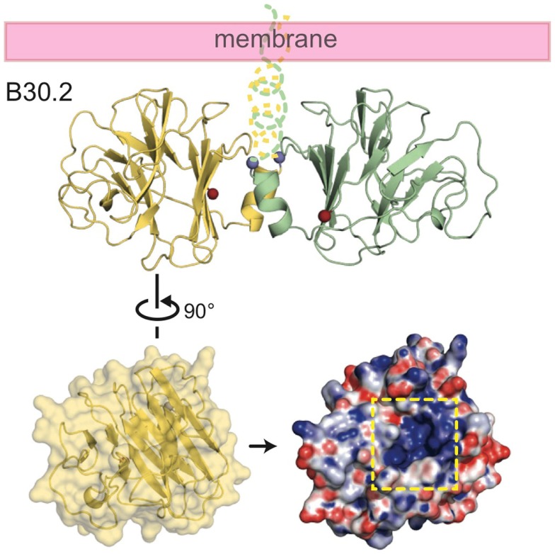
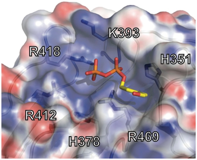
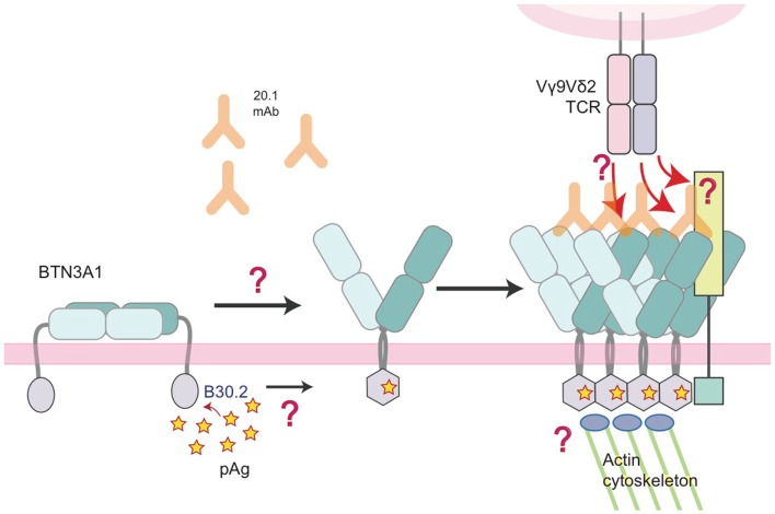
The predominant population of γδ T cells in human blood express a T cell receptor (TCR) composed of a Vγ9 (Vγ2 in an alternate nomenclature) and Vδ2 domains. These cells came into the limelight when it was discovered they can respond to certain microbial infections and tumorigenic cells through the detection of small, pyrophosphate containing organic molecules collectively called "phosphoantigens" or "pAgs." These molecules are intermediates in both eukaryotic and prokaryotic metabolic pathways. Chemical variants of these intermediates have been used in the clinic to treat a range of different cancers, however, directed optimization of these molecules requires a full understanding of their mechanism of action on target cells. We and others have identified a subclass of butyrophilin-related molecules (BTN3A1-3) that are directly involved in pAg sensing in the target cell, leading to engagement and activation of the T cell through the TCR. Our data and that of others support the pAg binding site to be the intracellular B30.2 domain of BTN3A1, which is the only isoform capable of mediating pAg-dependent stimulation of Vγ9Vδ2 T cells. Here, we review the data demonstrating pAg binding to the B30.2 domain and our studies of the structural conformations of the BTN3A extracellular domains. Finally, we synthesize a model linking binding of pAg to the intracellular domain with T cell detection via the extracellular domains in an "inside-out" signaling mechanism of the type characterized first for integrin molecule signaling. We also explore the role of Vγ9Vδ2 TCR variability in the CDR3 γ and δ loops and how this may modulate Vγ9Vδ2 cells as a population in surveillance of human health and disease.

Keywords: B30.2; T cell receptor; T cells; Vγ9Vδ2; butyrophilins; phosphoantigens.


Figures

Figure 1
Figure 1
Examples of phosphoantigens (pAgs) that stimulate Vγ9Vδ2 T cells. Phosphoantigens (pAgs) that derive from the mevalonate pathway (“endogenous” IPP and synthetic derivative EtPP) are circled in blue whereas those that produced in the isoprenoid pathway of microbes (“exogenous” HDMAPP or synthetic derivative cHDMAPP) are circled in pink. The pyrophosphate motif is highlighted in pink; the chemically diverse organic moieties are shown as lines.
Figure 2
Figure 2
Domain organization of the butryophilin-3 (BTN3) proteins. (A) The BTN3A extracellular domains are members of the B7-superfamily with a membrane-proximal IgC domain and an N-terminal IgV domain. The extracellular domains between the three isoforms are highly similar, but are linked, via a single-pass transmembrane region, to intracellular domains that vary amongst the three BTN3A family members. BTN3A1 and BTN3A3 both contain intracellular B30.2 domains whereas BTN3A2 does not. BTN3A1, circled with a dotted line, has been shown to be necessary for pAg-induced activation of Vγ9Vδ2 T cells. (B) The three-dimensional structures of the extracellular domain of BTN3A1 is shown in cyan (left) and superimposed with the A2 and A3 isoforms (right) shown in gold and pink, respectively. The structures are highly homologous, with only small variations in the hinge angles between the IgV and Ig-C domains.
Figure 3
Figure 3
Cartoon representation of the domain organization of the butryophilin-3 (BTN3) proteins. Structures of the extracellular domains of the BTN3A1 proteins shown in the two dimeric states present in the crystal lattice. Dimer 1 (left) associates via the IgC domains and forms a V-shaped dimer, placing the intracellular B30.2 domains in close proximity to each other. Dimer 2 (right) associates in an head-to-tail fashion with the IgV domain of one BTN3A monomer interacting with the IgC domain of another. This would result in the dimer laying parallel to the cell-surface, with the intracellular B30.2 domains separated. The interface contact residues are colored pink and shown on the surface representation of the two dimeric forms (middle panel). The buried surface area (BSA) is shown for both dimers.
Figure 4
Figure 4
Model of the regulation of BTN3A architecture by the agonist 20.1 and antagonist 103.2 antibodies. Structures of the extracellular domains of the BTN3A proteins (cyan) in complex with agonist (20.1, green) and antagonist (103.2, red) antibody single chains (scFv). The 20.1 antibody cannot “reach” across a BTN3A dimer 1 to occupy both binding sites and therefore is likely to multimerize BTN3A molecules on the cell-surface (left). The 20.1 antibody binds to the Dimer 2 interface of the IgV domain and therefore would disrupt the Dimer 2 conformation on the cell-surface. The 103.2 antibody can bind both Dimer 1 and Dimer 2 conformations, either potentially blocking the activating Dimer 1 form or stabilizing the “inactive” Dimer 2 form on the cell-surface.
Figure 5
Figure 5
Alignment of the intracellular B30.2 domains from BTN3A1 and BTN3A3. Amino acids are shown in the single letter designations, BTN3A1 is the consensus. “-” indicates positions of identity with BTN3A1, differences are shown in their single letter abbreviations. Green boxes indicate residues within 5 Å of the phosphoantigen binding pocket. BTN3A3 has an additional polypeptide extension.
Figure 6
Figure 6
Structure of the intracellular B30.2 domain of BTN3A1. Shown is a cartoon diagram of the B30.2 domain dimer identified in the crystal lattice. Monomer 1 is shown in yellow, monomer 2 in green. N- and C-termini are shown as blue and red spheres, respectively, as is the putative orientation of these molecules in relation to the cell membrane. Turning monomer 1 approximately 90° and generating an electrostatic representation of the B30.2 surface, a highly positively charged pocket is clear (indicated by the dashed yellow box).
Figure 7
Figure 7
Phosphoantigen binding pocket of B30.2 domain. Close-up view of the B30.2 pAg binding pocket with the side chains lining the pocket shown under the semi-transparent surface. The positions are labeled with the numbering relative to the full-length BTN3A1 molecule. The pAg is shown as sticks, modeled into the binding pocket; phosphates are colored orange and red (oxygen) and the organic moiety is shown in yellow.
Figure 8
Figure 8
Model of the molecular changes that occur in BTN3A molecules upon addition of 20.1 antibody or detection of accumulating intracellular pAg. In this model, we propose that BTN3A molecules exist on the surface of a healthy, unaffected cell in an inactive state, perhaps in a conformation similar to the dimer 2 visualized in the crystal lattice (left). Upon addition of the 20.1 antibody, dimer 2 is destabilized and BTN3A molecules are converted/stabilized in the dimer 1 conformation. An increase in the intracellular pAg concentration has a similar effect except the dimer conversion is mediated by changes in the intracellular B30.2 domain, which undergoes a conformational change upon pAg binding (hexagonal shape). This conformational change induces structural reorganization of BTN3A molecules, either via immobilization through B30.2 association with the actin cytoskeleton or B30.2 multimerization of BTN3A molecules, which is then signaled by an inside-out mechanism to change the architecture of the BTN3A extracellular domains. This architectural change could alone be the signal that Vγ9Vδ2 TCRs recognize, or recruitment of an additional, human-specific accessory molecule (lime-green square) occurs, which directly engages the Vγ9Vδ2 TCR.

References

    1. Salio M, Silk JD, Jones EY, Cerundolo V. Biology of CD1- and MR1-restricted T cells. Annu Rev Immunol (2014) 32:323–66.10.1146/annurev-immunol-032713-120243 - DOI - PubMed
    1. Born WK, Kemal Aydintug M, O’brien RL. Diversity of gammadelta T-cell antigens. Cell Mol Immunol (2013) 10:13–2010.1038/cmi.2012.45 - DOI - PMC - PubMed
    1. Chien YH, Meyer C, Bonneville M. Gammadelta T cells: first line of defense and beyond. Annu Rev Immunol (2014) 32:121–55.10.1146/annurev-immunol-032713-120216 - DOI - PubMed
    1. Kisielow J, Kopf M. The origin and fate of gammadelta T cell subsets. Curr Opin Immunol (2013) 25:181–8.10.1016/j.coi.2013.03.002 - DOI - PubMed
    1. Vantourout P, Hayday A. Six-of-the-best: unique contributions of gammadelta T cells to immunology. Nat Rev Immunol (2013) 13:88–100.10.1038/nri3384 - DOI - PMC - PubMed