Quantitative assessment of physiological cerebrospinal fluid flow in the cervical spinal canal with 3.0T phase-contrast cine MRI
- PMID: 25657672
- PMCID: PMC4308789
- DOI: 10.3969/j.issn.1673-5374.2012.18.005
Quantitative assessment of physiological cerebrospinal fluid flow in the cervical spinal canal with 3.0T phase-contrast cine MRI
Abstract
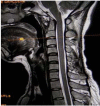
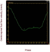
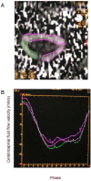
A total of 50 healthy volunteers aged between 18 and 54 years underwent phase-contrast cine MRI to assess cerebrospinal fluid flow characteristics in different regions of the vertebral canal. The results revealed that the cerebrospinal fluid peak flow velocity and peak flow rate in the systolic phase were significantly greater than those in the diastolic phase at the same level in the subarachnoid space of the cervical spinal canal. The ventral peak flow velocity and peak flow rate were significantly greater than the post-lateral peak flow velocity and flow rate, while there were no differences between left and right post-lateral subarachnoid peak velocity and flow rate. Moreover, there were no significant differences in peak flow velocity and peak flow rate between the systolic and diastolic phases, ventral, right post-lateral or left post-lateral peak flow velocity and peak flow rate at the same level in the subarachnoid space of the cervical spinal canal among different age groups (18-24, 25-34, 35-44, ≥ 45 years).
Keywords: cerebrospinal fluid; flow velocity; magnetic resonance imaging; neural regeneration; phase-contrast; subarachnoid space; vertebral canal.
Conflict of interest statement
Figures


Similar articles
-
Quantitative analysis of intraspinal cerebrospinal fluid flow in normal adults.Neural Regen Res. 2012 May 25;7(15):1164-9. doi: 10.3969/j.issn.1673-5374.2012.15.007. Neural Regen Res. 2012. PMID: 25722710 Free PMC article.
-
Quantitative analysis of cerebrospinal fluid flow in patients with cervical spondylosis using cine phase-contrast magnetic resonance imaging.Neurosurgery. 1999 Apr;44(4):779-84. doi: 10.1097/00006123-199904000-00052. Neurosurgery. 1999. PMID: 10201303
-
Characterising spinal cerebrospinal fluid flow in the pig with phase-contrast magnetic resonance imaging.Fluids Barriers CNS. 2023 Jan 18;20(1):5. doi: 10.1186/s12987-022-00401-4. Fluids Barriers CNS. 2023. PMID: 36653870 Free PMC article.
-
Elucidating the pathophysiology of syringomyelia.J Neurosurg. 1999 Oct;91(4):553-62. doi: 10.3171/jns.1999.91.4.0553. J Neurosurg. 1999. PMID: 10507374
-
Quantitative assessment of surgical decompression of the cervical spine with cine phase contrast magnetic resonance imaging.Neurosurgery. 2002 Apr;50(4):791-5; discussion 796. doi: 10.1097/00006123-200204000-00020. Neurosurgery. 2002. PMID: 11904030
References
-
- O’Connel JEA. The vascular factor tn intracranial pressure and the maintenance of the cerebrospinal fluid circulation. Brain. 1943;66(3):204–228.
-
- Bradley WG, Jr, Whittemore AR, Kortman KE, et al. Marked cerebrospinal fluid void: indicator of successful shunt in patients with suspected normal-pressure hydrocephalus. Radiology. 1991;178(2):459–466. - PubMed
-
- Moran PR. A flow velocity zeugmatographic interlace for NMR imaging in humans. Magn Reson Imaging. 1982;1(4):197–203. - PubMed
-
- Mascalchi M, Ciraolo L, Tanfani G, et al. Cardiac-gated phase MR imaging of aqueductal CSF flow. J Comput Assist Tomogr. 1988;12(6):923–926. - PubMed
-
- Penn RD, Basati S, Sweetman B, et al. Ventricle wall movements and cerebrospinal fluid flow in hydrocephalus. J Neurosurg. 2011;115(1):159–164. - PubMed
LinkOut - more resources
Full Text Sources
