

Improved C3-4 transfer for treatment of root avulsion of the brachial plexus upper trunk: Animal experiments and clinical application
- PMID: 25657692
- PMCID: PMC4308750
- DOI: 10.3969/j.issn.1673-5374.2012.20.004
Improved C3-4 transfer for treatment of root avulsion of the brachial plexus upper trunk: Animal experiments and clinical application
Abstract
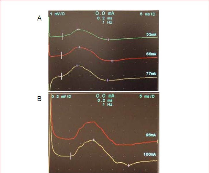
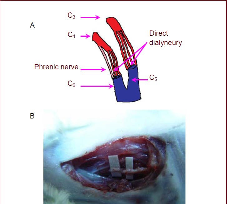
Experimental rats with root avulsion of the brachial plexus upper trunk were treated with the improved C3-4 transfer for neurotization of C5-6. Results showed that Terzis grooming test scores were significantly increased at 6 months after treatment, the latency of C5-6 motor evoked potential was gradually shortened, and the amplitude was gradually increased. The rate of C3 instead of C5 and the C4 + phrenic nerve instead of C6 myelinated nerve fibers crossing through the anastomotic stoma was approximately 80%. Myelinated nerve fibers were arranged loosely but the thickness of the myelin sheath was similar to that of the healthy side. In clinical applications, 39 patients with root avulsion of the brachial plexus upper trunk were followed for 6 months to 4.5 years after treatment using the improved C3 instead of C5 nerve root transfer and C4 nerve root and phrenic nerve instead of C6 nerve root transfer. Results showed that the strength of the brachial biceps and deltoid muscles recovered to level III-IV, scapular muscle to level III-IV, latissimus dorsi and pectoralis major muscles to above level III, and the brachial triceps muscle to level 0-III. Results showed that the improved C3-4 transfer for root avulsion of the brachial plexus upper trunk in animal models is similar to clinical findings and that C3-4 and the phrenic nerve transfer for neurotization of C5-6 can innervate the avulsed brachial plexus upper trunk and promote the recovery of nerve function in the upper extremity.
Keywords: brachial plexus; cervical plexus; nerve transfer; neural regeneration; peripheral nerve injury; phrenic nerve; root avulsion; translational medicine; upper trunk.
Conflict of interest statement
Figures


Similar articles
-
[An experimental study on outcome of ipsilateral C7 nerve root transfer to repair the root avulsion of the brachial plexus].Zhonghua Wai Ke Za Zhi. 2008 May 15;46(10):763-7. Zhonghua Wai Ke Za Zhi. 2008. PMID: 18953933 Chinese.
-
Effect of ipsilateral C7 nerve root transfer on restoration of rat upper trunk muscle and nerve function after brachial plexus root avulsion.Orthopedics. 2010 Dec 1;33(12):886. doi: 10.3928/01477447-20101021-12. Orthopedics. 2010. PMID: 21162507
-
Repair of brachial plexus lesions by end-to-side side-to-side grafting neurorrhaphy: experience based on 11 cases.Microsurgery. 2005;25(2):126-46. doi: 10.1002/micr.20036. Microsurgery. 2005. PMID: 15389968
-
[Paralytic shoulder secondary to post-traumatic peripheral nerve lesions in the adult].Acta Orthop Belg. 1999 Mar;65(1):10-22. Acta Orthop Belg. 1999. PMID: 10216997 Review. French.
-
[Recent progress in diagnosis and treatment of the injury to the peripheral nerve].Zhongguo Xiu Fu Chong Jian Wai Ke Za Zhi. 2006 Apr;20(4):319-23. Zhongguo Xiu Fu Chong Jian Wai Ke Za Zhi. 2006. PMID: 16683422 Review. Chinese.
Cited by
-
Partial Recovery of Limb Function Following End-to-Side Screw Anastomosis of Phrenic Nerve in Rats with Brachial Plexus Injury.Med Sci Monit. 2018 Jul 12;24:4832-4840. doi: 10.12659/MSM.908379. Med Sci Monit. 2018. PMID: 30001299 Free PMC article.
References
-
- Tsuyama N, Hara T. Intercostal nerve transfer in the treatment of brachial plexus injury of root avulsion type. In: Delchef J, de Mameffe R, Vander Elst E, editors. Orthopaedic Surgery and Traumatology. Amesterdam: International Congress Series No. 291, Excerpta Medica; 1973. pp. 351–353.
-
- Gu YD, Wu MM, Zheng YL, et al. Phrenic nerve transfer treating root avulsion of the brachial plexus. Zhonghua Wai Ke Za Zhi. 1989;27(7):433–435. 447. - PubMed
-
- Oberlin C, Béal D, Leechavengvongs S, et al. Nerve transfer to biceps muscle using a part of ulnar nerve for C5-C6 avulsion of the brachial plexus: anatomical study and report of four cases. J Hand Surg Am. 1994;19(2):232–237. - PubMed
-
- Gu YD, Zhang GM, Chen DS, et al. Cervical nerve root transfer from the healthy side in the treatment of brachial plexus root avulsion. Zhonghua Yi Xue Za Zhi. 1989;69(10):563–538. - PubMed
-
- Xu JG, Hu SN, Wang H, et al. Histochemical study on C7 roots and its clinical significance. Zhongguo Linchuang Jiepou Xue Zazhi. 1996;14(4):243–245.
LinkOut - more resources
Full Text Sources
Research Materials
Miscellaneous