Review
. 2014 Dec 1;9(23):2048-52.
doi: 10.4103/1673-5374.147929.

Dynamic reactive astrocytes after focal ischemia

Affiliations
Review

Dynamic reactive astrocytes after focal ischemia

Shinghua Ding. Neural Regen Res. .

Abstract

Astrocytes are specialized and most numerous glial cell type in the central nervous system and play important roles in physiology. Astrocytes are also critically involved in many neural disorders including focal ischemic stroke, a leading cause of brain injury and human death. One of the prominent pathological features of focal ischemic stroke is reactive astrogliosis and glial scar formation associated with morphological changes and proliferation. This review paper discusses the recent advances in spatial and temporal dynamics of morphology and proliferation of reactive astrocytes after ischemic stroke based on results from experimental animal studies. As reactive astrocytes exhibit stem cell-like properties, knowledge of dynamics of reactive astrocytes and glial scar formation will provide important insights for astrocyte-based cell therapy in stroke.

Keywords: cell proliferation; cell therapy; dynamics; glial scar; ischemic stroke; morphology; reactive astrocytes.


Figures
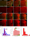
Figure 1
Figure 1
Time course of astrocyte proliferation and morphological changes after stroke. (A) Fluorescent images of glial fibrillary acidic protein (GFAP) and BrdU expression in the penumbra from mice at different times after PT. (B) Summary of bromodeoxyuridine (BrdU) + cell density presented as cell number per mm2 in region 1 (R1) and region (R2) with an area of 200 μm × 200 μm in the layers 2/3 of cortex. R1 is located 0–200 μm from the edge of ischemic core, and R2 is located 200–400 μm from the edge of ischemic core (see the left panel of A2). (C, D) The density of GFAP+ (C) and GFAP+BrdU+ (D) stained cells in the penumbra. The cells were counted in the penumbral region of 200 μm × 400 μm in layers 2/3 cortex located 0–400 μm from the edge of ischemic core. IC: Ischemic core; P: penumbra. Data were adapted from Li et al. (2014).
Figure 2
Figure 2
Schematic representations of dynamic reactive astrocytes in the penumbra and glial scar formation at different stages after a focal ischemic stroke. (A) In control conditions, very low percentage of astrocytes expresses glial fibrillary acidic protein (GFAP). (B) Acute phase after focal ischemia (days 1–4 post ischemia). Astrocytes exhibit stellate morphology and hypertrophied GFAP positive processes and a high proliferating rate. (C) Sub-acute phase after focal ischemia (days 4–8 post ischemia). Astrocytes exhibit elongated processes pointing to the ischemic core, and a glial scar is formed. The pro-liferating rate decreases significantly at this stage. (D) Chronic phase after focal ischemia (longer than day 8 post ischemia). Astrocytes further extend processes toward the ischemic core and glial scar is matured. The GFAP expression levels in reactive astrocytes surrounding the glial scar decreased and reactive astrocytes lose the capability of proliferation. Astrocytes are heterogeneous in morphology, molecular expression, and proliferation.

References

    1. Addis RC, Hsu FC, Wright RL, Dichter MA, Coulter DA, Gearhart JD. Efficient conversion of astrocytes to functional midbrain dopaminergic neurons using a single polycistronic vector. PLoS One. 2011;6:e28719. - PMC - PubMed
    1. Badan I, Buchhold B, Hamm A, Gratz M, Walker LC, Platt D, Kessler C, Popa-Wagner A. Accelerated glial reactivity to stroke in aged rats correlates with reduced functional recovery. J Cereb Blood Flow Metab. 2003;23:845–854. - PubMed
    1. Bao Y, Qin L, Kim E, Bhosle S, Guo H, Febbraio M, Haskew-Layton RE, Ratan R, Cho S. CD36 is involved in astrocyte activation and astroglial scar formation. J Cereb Blood Flow Metab. 2012;32:1567–1577. - PMC - PubMed
    1. Barreto GE, Sun X, Xu L, Giffard RG. Astrocyte proliferation following stroke in the mouse depends on distance from the Infarct. PLoS One. 2011;6:e27881. - PMC - PubMed
    1. Benesova J, Hock M, Butenko O, Prajerova I, Anderova M, Chvatal A. Quantification of astrocyte volume changes during ischemia in situ reveals two populations of astrocytes in the cortex of GFAP/EGFP mice. J Neurosci Res. 2009;87:96–111. - PubMed