

doi: 10.4103/1673-5374.147942.
A novel mode of retinal regeneration: the merit of a new Xenopus model
Affiliations
- PMID: 25657732
- PMCID: PMC4316444
- DOI: 10.4103/1673-5374.147942
Item in Clipboard
A novel mode of retinal regeneration: the merit of a new Xenopus model
Neural Regen Res.
.
No abstract available
Figures
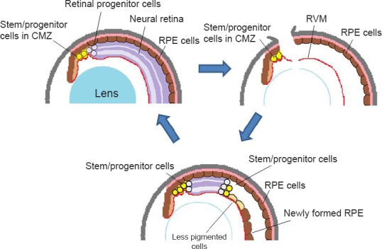
A schema for retinal regeneration in X. tropicalis: stem/progenitor cells in the ciliary marginal zone regenerate the whole retina. (A) Normal eye. During the growth of the eye, stem/progenitor cells in the ciliary marginal zone (CMZ) produce retinal progenitor cells, which are incorporated into the retina at the periphery. (B) An eye immediately after retinal removal. An incision is made at the dorsal periphery of the eye, and the lens fibers are removed, leaving the retinal vascular membrane (RVM) in the posterior chamber. Some of the retinal pigmented epithelial (RPE) cells begin to be detached and migrate to the RVM, where they form a new RPE layer (as seen in Figure C). (C) At the later stage of regeneration (days 15–20), the regenerating retina extends further to the posterior. At the posterior edge of the regenerating retina, a proliferating zone (progenitor zone) is observed (consisting of actively proliferating cells). At the peripheral end of the regenerating retina (closer to the iris) there still remain progenitor cells that are intensely labeled for BrdU. The regenerating retina is sandwiched by progenitor cell zones at both ends. In X. tropicalis, RPE cells in the newly formed epithelium do not appear to transdifferentiate into neuronal cells, although some cells at the boundary to the regenerating retina become less pigmented. These cells are gradually lost as the regenerating retina extends more posteriorly. Contrary to X. tropicalis, the RPE cells on the RVM transdifferentiate into the retina in X. laevis. The schema is modified from the original figure (Miyake and Araki, 2014).
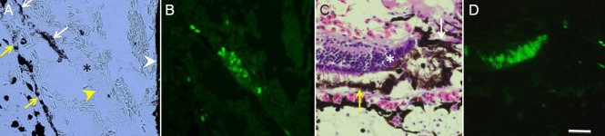
BrdU labeling of stem/progenitor cells at the early stage of regeneration (day 5) (A, B) and late stage (day 20) (C, D). (A, B) At the early stage, there is an actively proliferating cell zone (asterisk in A) juxtaposed to the presumable ciliary marginal zone (CMZ) (yellow arrowhead) and the iris (white arrowhead). This proliferating cell zone (progenitor cells) appear continuous to the newly formed retinal pigmented epithelial (RPE) layer (white arrow). The original RPE layer is shown by yellow arrows. A is a phase contrast micrograph image and is identical to B. (C, D) At the later stage, there is another progenitor cell clusters (asterisk) at the posterior end of regenerating retina. There still remain some RPE cells (white arrow). The original RPE is shown by a yellow arrow. C is a micrograph of a section stained with hematoxylin and eosin, and is identical to D. Scale bar in D is 45 μm and applied to A–C.
Comment on
- Neural Regen Res. 9(24):2122.
References
-
- Araki M. A model for retinal regeneration in Xenopus. In ‘Xenopus Development’. In: Kloc M, Kubiak J Z, editors. John Wiley & Sons, Inc; 2014. pp. 346–367.
-
- Borday C, Cabochette P, Parain K, Mazurier N, Janssens S, Tran HT, Sekkali B, Bronchain O, Vleminckx K, Locker M, Perron M. Antagonistic cross-regulation between Wnt and Hedgehog signalling pathways controls post-embryonic retinal proliferation. Development. 2012;139:3499–509. - PubMed
-
- Chiba C, Mitashov VI. Cellular and molecular events in the adult newt retinal regeneration. In: Chiba C, editor. “Strategies for retinal tissue repair and regeneration in vertebrantes: From fish to human”. Kerala, India: Research Signpost; 2007. pp. 16–37.
-
- DelRio-Tsonis K, Tsonis PA. Eye regeneration at the molecular age. Dev Dyn. 2003;226:211–224. - PubMed
-
- Fisher AJ. Neural regeneration in the chick retina. Prog Retina Eye Res. 2005;24:161–182. - PubMed
LinkOut - more resources
Full Text Sources
Other Literature Sources
Research Materials