

The human trochlear and abducens nerves at different ages - a morphometric study
- PMID: 25657848
- PMCID: PMC4306475
- DOI: 10.14336/AD.2014.0310
The human trochlear and abducens nerves at different ages - a morphometric study
Abstract
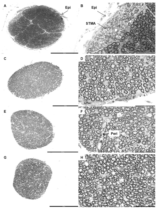
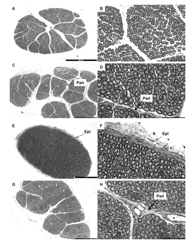
The trochlear and abducens nerves (TN and AN) control the movement of the superior oblique and lateral rectus muscles of the eyeball, respectively. Despite their immense clinical and radiological importance no morphometric data was available from a wide spectrum of age groups for comparison with either pathological or other conditions involving these nerves. In the present study, morphometry of the TN and AN was performed on twenty post-mortem samples ranging from 12-90 years of age. The nerve samples were processed for resin embedding and toluidine blue stained thin (1µm) sections were used for estimating the total number of myelinated axons by fractionator and the cross sectional area of the nerve and the axons by point counting methods. We observed that the TN was covered by a well-defined epineurium and had ill-defined fascicles, whereas the AN had multiple fascicles with scanty epineurium. Both nerves contained myelinated and unmyelinated fibers of various sizes intermingled with each other. Out of the four age groups (12-20y, 21-40y, 41-60y and >61y) the younger groups revealed isolated bundles of small thinly myelinated axons. The total number of myelinated fibers in the TN and AN at various ages ranged from 1100-3000 and 1600-7000, respectively. There was no significant change in the cross-sectional area of the nerves or the axonal area of the myelinated nerves across the age groups. However, myelin thickness increased significantly in the AN with aging (one way ANOVA). The present study provides baseline morphometric data on the human TN and AN at various ages.
Keywords: morphometry; myelin thickness; ocular motor nerves; stereology.
Figures
References
-
- Glimcher PW. Eye movements. In: Squire LR, Bloom FE, McConnel SK, Roberts JL, Spitzer NC, Zigmond MJ, editors. Fundamental Neuroscience. 2nd Edn. New York: Academic Press; 2003. pp. 873–892.
-
- Calisaneller T, Ozdemir O, Altinors N. Posttraumatic acute bilateral abducens nerve palsy in a child. Childs Nerv Syst. 2006;22:726–728. - PubMed
-
- Dwarakanath S, Gopal S, Venkataramana NK. Post-traumatic bilateral abducens nerve palsy. Neurol India. 2006;54:221–222. - PubMed
-
- Kurbanyan K, Lessell S. Intracranial hypotension and abducens palsy following upper spinal manipulation. Br J Ophthalmol. 2008;92:153–155. - PubMed
-
- Hanu-Cernat LM, Hall T. Late onset of abducens palsy after Le Fort I maxillary osteotomy. Br J Oral Maxillofac Surg. 2009;47:414–416. - PubMed
LinkOut - more resources
Full Text Sources