

Spatial mapping of proteoglycan content in articular cartilage using near-infrared (NIR) spectroscopy
- PMID: 25657883
- PMCID: PMC4317110
- DOI: 10.1364/BOE.6.000144
Spatial mapping of proteoglycan content in articular cartilage using near-infrared (NIR) spectroscopy
Abstract
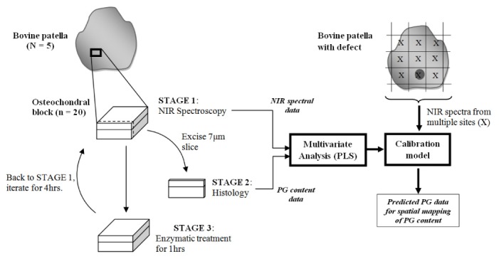
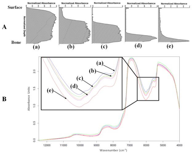
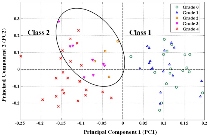
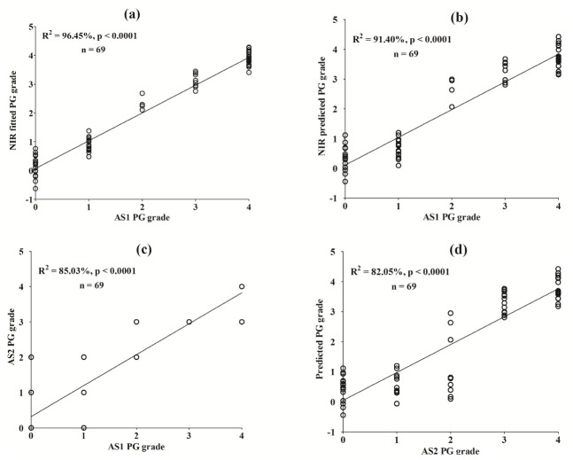
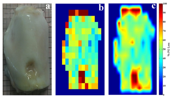
Diagnosis of articular cartilage pathology in the early disease stages using current clinical diagnostic imaging modalities is challenging, particularly because there is often no visible change in the tissue surface and matrix content, such as proteoglycans (PG). In this study, we propose the use of near infrared (NIR) spectroscopy to spatially map PG content in articular cartilage. The relationship between NIR spectra and reference data (PG content) obtained from histology of normal and artificially induced PG-depleted cartilage samples was investigated using principal component (PC) and partial least squares (PLS) regression analyses. Significant correlation was obtained between both data (R(2) = 91.40%, p<0.0001). The resulting correlation was used to predict PG content from spectra acquired from whole joint sample, this was then employed to spatially map this component of cartilage across the intact sample. We conclude that NIR spectroscopy is a feasible tool for evaluating cartilage contents and mapping their distribution across mammalian joint.
Keywords: (170.3880) Medical and biological imaging; (170.6510) Spectroscopy, tissue diagnostics; (170.6935) Tissue characterization.
Figures
References
-
- Buckwalter J. A., Mankin H. J., “Articular Cartilage: Degeneration and Osteoarthrosis, Repair, Regeneration, and Transplantation,” J. Bone Jt. Surg. 47, 612–632 (1998). - PubMed
-
- Saarakkala S., Julkunen P., Kiviranta P., Mäkitalo J., Jurvelin J. S., Korhonen R. K., “Depth-wise progression of osteoarthritis in human articular cartilage: investigation of composition, structure and biomechanics,” Osteoarthritis Cartilage 18(1), 73–81 (2010). 10.1016/j.joca.2009.08.003 - DOI - PubMed
LinkOut - more resources
Full Text Sources
Other Literature Sources
Miscellaneous