Beta-catenin expression in psoriasis
- PMID: 25657910
- PMCID: PMC4314880
- DOI: 10.4103/2229-5178.148923
Beta-catenin expression in psoriasis
Abstract
Background: Psoriasis is a common inflammatory skin disease characterized by abnormal keratinocyte proliferation and differentiation. Beta-catenin participates in intercellular adhesion. Catenins are proteins found in complexes with cadherin cell adhesion molecules of cells. The role of catenin in regulating keratinocyte stem cell differentiation and hair follicle morphogenesis has been extensively reported.
Aims and objectives: is to study β-catenin expression in lesional and non-lesional psoriatic skin to throw light upon its possible role in the pathogenesis of psoriasis.
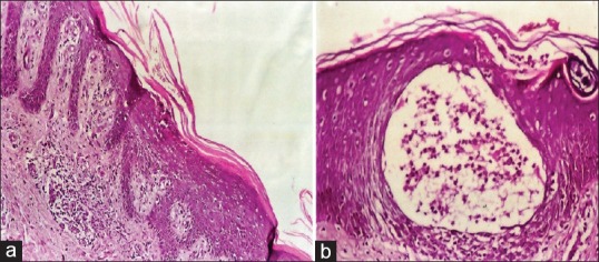
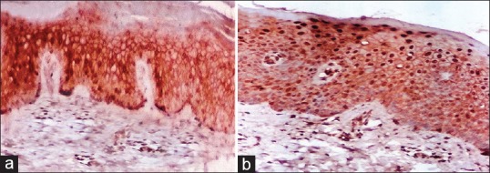
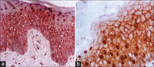
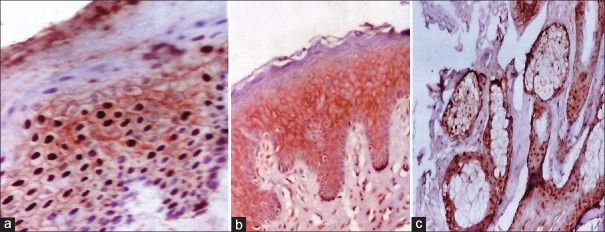
Materials and methods: Biopsies were taken from 20 patients with psoriasis vulgaris and from 10 normal controls. The distribution of Beta catenin was investigated using polycolonal rabbits B-catenin antibody-1 by immunohistochemical method.
Results: In this study membranous β-catenin expression was significantly demonstrated in the control group then the non-lesional areas in comparison to the lesional areas (P < 0.001). Nuclear β-catenin staining expression was significantly more demonstrated in lesional and non-lesional areas in comparison to the control cases (P < 0.001).
Conclusions: The down regulation of membranous β-catenin expression in lesional psoriatic skin might reflect a useful phenotypic marker of hyperprolifration of keratinocytes in psoriasis. Moreover, the mild down regulation of membranous β-catenin expression in non lesional psoriatic skin may provide clues about incipient structural abnormalities in the pathogenesis of psoriasis, providing an early diagnostic indicator for evolution to a generalized form of the disease. Nuclear β-catenin expression was not found in the control group but was demonstrated in lesional and moderately in non-lesional reflecting its role in kerationcyte proliferation.
Keywords: Beta-catenin; pathogenesis-immunohistochemical; psoriasis.
Conflict of interest statement
Figures
Similar articles
-
Increased nuclear beta-catenin in suprabasal involved psoriatic epidermis.Br J Dermatol. 2007 Dec;157(6):1168-77. doi: 10.1111/j.1365-2133.2007.08195.x. Epub 2007 Oct 4. Br J Dermatol. 2007. PMID: 17916213 Clinical Trial.
-
Progranulin and beta-catenin in psoriasis: An immunohistochemical study.J Cosmet Dermatol. 2019 Dec;18(6):2019-2026. doi: 10.1111/jocd.12966. Epub 2019 May 15. J Cosmet Dermatol. 2019. PMID: 31091001
-
Expression of p53 protein in psoriasis.Acta Dermatovenerol Alp Pannonica Adriat. 2005 Sep;14(3):79-83. Acta Dermatovenerol Alp Pannonica Adriat. 2005. PMID: 16200332
-
Role of CYR61 in psoriatic lesional and perilesional skin: A clinical and immunohistochemical study.J Cosmet Dermatol. 2021 Sep;20(9):2981-2988. doi: 10.1111/jocd.13947. Epub 2021 Jan 22. J Cosmet Dermatol. 2021. PMID: 33484099
-
Intralesional T-lymphocyte activation as a mediator of psoriatic epidermal hyperplasia.J Invest Dermatol. 1995 Jul;105(1 Suppl):89S-94S. doi: 10.1111/1523-1747.ep12316121. J Invest Dermatol. 1995. PMID: 7616005 Review.
Cited by
-
A Case of Acute Generalized Pustular Psoriasis of von Zumbusch Triggered by Hypocalcemia.Case Rep Dermatol. 2015 Dec 3;7(3):345-51. doi: 10.1159/000442380. eCollection 2015 Sep-Dec. Case Rep Dermatol. 2015. PMID: 26955330 Free PMC article.
-
PGRN Suppresses Inflammation and Promotes Autophagy in Keratinocytes Through the Wnt/β-Catenin Signaling Pathway.Inflammation. 2016 Aug;39(4):1387-94. doi: 10.1007/s10753-016-0370-y. Inflammation. 2016. PMID: 27239673
-
Using Imiquimod-Induced Psoriasis-Like Skin as a Model to Measure the Skin Penetration of Anti-Psoriatic Drugs.PLoS One. 2015 Sep 10;10(9):e0137890. doi: 10.1371/journal.pone.0137890. eCollection 2015. PLoS One. 2015. PMID: 26355594 Free PMC article.
-
IL-36γ inhibits differentiation and induces inflammation of keratinocyte via Wnt signaling pathway in psoriasis.Int J Med Sci. 2017 Aug 18;14(10):1002-1007. doi: 10.7150/ijms.20809. eCollection 2017. Int J Med Sci. 2017. PMID: 28924372 Free PMC article.
-
Changes in Proteome of Fibroblasts Isolated from Psoriatic Skin Lesions.Int J Mol Sci. 2020 Jul 28;21(15):5363. doi: 10.3390/ijms21155363. Int J Mol Sci. 2020. PMID: 32731552 Free PMC article.
References
-
- Louden BA, Pearce DJ, Lang W, Feldman SR. A Simplified Psoriasis Area Severity Index (SPASI) for rating psoriasis severity in clinic patients. Dermatol Online J. 2004;10:7. - PubMed
-
- Ghorpade A. Linear naevoid psoriasis along lines of Blaschko. J Eur Acad Dermatol Venereol. 2004;18:726–7. - PubMed
-
- Eckert RL, Sturniolo MT, Broome AM, Ruse M, Rorke EA. Transglutaminase function in epidermis. J Invest Dermatol. 2005;124:481–92. - PubMed
-
- Iizuka H, Takahashi H, Honma M, Ishida-Yamamoto A. Unique keratinization process in psoriasis: Late differentiation markers are abolished because of the premature cell death. J Dermatol. 2004;31:271–6. - PubMed
LinkOut - more resources
Full Text Sources
Other Literature Sources