. 2015 Feb 6;10(2):e0117247.
doi: 10.1371/journal.pone.0117247. eCollection 2015.

The host-protective effect of arabinosylated lipoarabinomannan against Leishmania donovani infection is associated with restoration of IFN-γ responsiveness

Affiliations

The host-protective effect of arabinosylated lipoarabinomannan against Leishmania donovani infection is associated with restoration of IFN-γ responsiveness

Bidisha Paul Chowdhury et al. PLoS One. .

Abstract

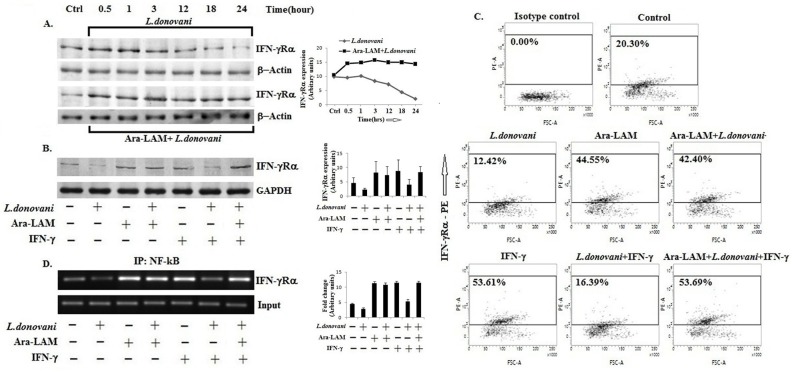
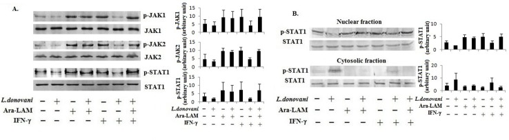
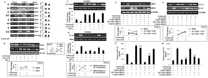
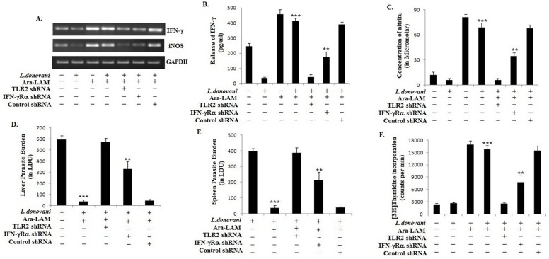
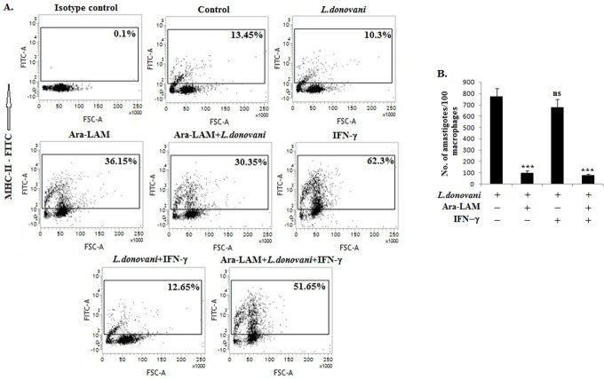
Visceral leishmaniasis (VL), which is endemic as a major infectious disease in the tropical and subtropical countries, is caused by a protozoan parasite Leishmania donovani. At present, restricted treatment options and lack of vaccines intensify the problem of controlling VL. Therefore, finding a novel immunoprophylactic or therapeutic principle is a pressing need. Here, we report that arabinosylated lipoarabinomannan (Ara-LAM), a TLR2-ligand isolated from Mycobacterium smegmatis, exhibits a strong immunomodulatory property that conferred protection against L. donovani infection. Although, Ara-LAM modulates TLR2 and MAPK signaling, it is not known whether Ara-LAM involves IFN-γ signaling for effective parasite clearance. Because, it is reported that IFN-γ signaling, a principle mediator of NO generation and macrophage and Tcell activation, is hampered during leishmanial pathogenesis. Ara-LAM increases IFN-γ receptor expression and potentiates IFN-γ receptor signaling through JAK-STAT pathway. Moreover, Ara-LAM reciprocally modulates IRF4 and IRF8 expression and reinstates anti-leishmanial Th1 response that eventuates in significantly reduced parasite load in spleen and liver of L. donovani-infected BALB/c mice. IFN-γRα silencing resulted in the suppression of these host-protective mechanisms affected by Ara-LAM. Thus, Ara-LAM-mediated restoration of IFN-γ responsiveness is a novel immuno-modulatory principle for protection against L. donovani susceptible host.


Conflict of interest statement

Competing Interests: The authors have declared that no competing interests exist.

Figures

Fig 1
Fig 1. Ara-LAM enhanced IFN-γ Receptor alpha chain expression in parasitized macrophages.
(A) BALB/c derived peritoneal macrophages (2x10⁶cells) were infected with Leishmania donovani (cell: parasite 1:10) for 30mins, 1hr, 3hrs, 12hrs, 18hrs and 24hrs. Other sets of macrophages were pre-treated with Ara-LAM for 3 hrs and followed by infection for the same time periods. Cell lysates were prepared followed by Western blot for IFN-γRα. Data are from 1 of 3 experiments conducted in the same way with similar results. IFN-γRα expression in Ara-LAM treated infected macrophages were significantly higher than the infected cells (p<0.05). (B) In a separate set of experiment, BALB/c derived peritoneal macrophages (2x10⁶cells) were pre-treated with Ara-LAM for 3 hr and followed by Leishmania donovani infection for 24 hr. Macrophages were then treated with rIFN-γ (20ng/ml) for 45min, followed by cell lysate preparation and Western blotting for IFN-γRα. The blots shown are representative of three experiments. (C) Ara-LAM pre-treated peritoneal macrophages (2x10⁶cells) were infected with L. donovani for 24 hrs followed by rIFN-γ (20ng/ml) stimulation for another 24 hrs, and staining with IFN-γRα-PE Ab and analyzed on a flow cytometer. Data are from one representative experiment that was repeated thrice. (D) Treated and untreated infected macrophages (2x10⁶cells) were subjected to immuno-precipitation using NF-κB (IP:NF-κB) specific Ab. Semi quantitative RT-PCR was performed for amplifying the putative NF-κB binding sites at the IFN-γRα promoter. Data are from one representative experiment, which was performed at least thrice.
Fig 2
Fig 2. Moderation of IFN-γ induced JAK-STAT signaling by Ara-LAM in Leishmania infected macrophages.
(A) BALB/c derived peritoneal macrophages were pre-treated with Ara-LAM for 3 hrs, followed by L. donovani infection for 24hrs. Macrophages were treated with rIFN-γ (20ng/ml) for 45min, followed by cell lysate preparation and Western blot for JAK1, p-JAK1, JAK2, p-JAK2, STAT-1 and p-STAT-1. The blots shown are representative of triplicate experiments. (B) Both the nuclear extracts and the cytosolic extracts of differently treated peritoneal macrophages were prepared followed by Western blot to analyze the nuclear translocation of p-STAT-1 in L. donovani infected macrophages. Blots shown here are from one of three representative experiments.
Fig 3
Fig 3. Ara-LAM reciprocally regulated IRF4 and IRF8 expression and corresponding immune response during L. donovani infection both in vitro and in vivo.
(A) L. donovani infected and uninfected control peritoneal macrophages were collected in Trizol for RNA extraction and semi-quantitative RT-PCR was performed. Data represent means ± SD for three experiments. Inset 1: Comparison between IRF4 and IRF8 expression in L. donovani infected and uninfected control macrophages. (B) Peritoneal macrophages (2x10⁶cells) from BALB/c mice were cultured and subjected to Ara-LAM pre-treatment (3μg/ml) for 3hrs, followed by L. donovani challenge for 6hrs. Both rIFN-γ stimulated (20ng/ml, 45mins) and unstimulated cells were collected in Trizol for RNA extraction and semi-quantitative RT-PCR was performed. Data are from one representative experiment, which was performed at least thrice. Inset 2: Reciprocal expression of IRF4 and IRF8 by Ara-LAM in L. donovani infected macrophages. (C-D) BALB/c derived peritoneal macrophages (2x10⁶cells) were treated with Ara-LAM (3μg/ml) for 3hrs, followed by L. donovani challenge for 6hrs. After 45 min of rIFN-γ stimulation immuno-precipitations were conducted using IRF8 (IP:IRF8) and IRF4 (IP: IRF4) specific Abs. Semi quantitative RT-PCR was performed for amplifying the putative IRF8 binding sites of the IL-12p40 promoter and IRF4 binding sites of the IL-10 promoter. Data represent means ± SD for three sets of experiments. Inset 3: Ara-LAM mediated up-regulation of IRF8 binding to the IL-12 promoter and down-regulation of IRF4 binding to the IL-10 promoter in infected macrophages. (E-F) BALB/c mice were injected with respective shRNAs (for 2 days) followed by treatment with either phosphate-buffered saline (PBS) (control) or Ara-LAM (30 μg intraperitoneally) for 2 days, after which mice were infected. 28 days later, mice were sacrificed and splenocytes (2x10⁶) were collected in Trizol for mRNA extraction and semi-quantitative reverse transcriptase polymerase chain reaction (RT-PCR) analysis (see Methods). Data are from one of three representative experiments. Inset 4: Correlations (R-values) between IRF4 and IL-10 or IL-12 expression in Ara-LAM treated and untreated infected splenocytes. Inset 5: Correlations (R-values) between IRF8 and IL-10 or IL-12 expression in same set of mice. (G-H) A separate set of splenocytes (2x10⁶) were stimulated with soluble leishmanial antigen (SLA) at 5 μg/ml for 48 h. Release of Interleukin 12 (IL-12) p70 and Interleukin 10 (IL-10) culture supernatants were determined by enzyme-linked immunosorbent assay. Data represent means ± SD for 4 animals per group. ***P <.001 and **P < .01 for the comparison with infected mice.
Fig 4
Fig 4. Effect of Ara-LAM and IFN-γ signaling against leishmanial pathogenesis in BALB/c mice.
(A) Mice were injected with respective shRNAs (for 2 days) followed by treatment with either phosphate-buffered saline (PBS) (control) or Ara-LAM (30 μg intraperitoneally) for 2 days, after which mice were infected. After 28 days mice were sacrificed, splenocytes (2x10⁶) was collected in Trizol for mRNA extraction and semi-quantitative reverse transcriptase polymerase chain reaction (RT-PCR) analysis (see Methods). Data are from one of three representative experiment. (B-C) A separate set of splenocytes (2x10⁵cells/well) was stimulated with soluble leishmanial antigen (SLA) at 5 μg/ml for 48 h. Release of Interferon-γ (IFN-γ) and nitric oxide in culture supernatants were determined by enzyme-linked immunosorbent assay and the Nitric Oxide Colorimetric Assay kit, respectively. Data represent means ± SD for four animals per group. ***P <.001 and **P <. 01 for the comparison with infected mice. (D-E) Differently treated infected mice were sacrificed 28 days after infection. Levels of parasite burden in liver and spleen are expressed in Leishman-Donovan units (LDUs). Data represent means ± SD for 4 animals per group. ***P <.001 and **P < .01 for the comparison with infected mice. (F) Proliferative responses to soluble leishmanial antigen (SLA) (5 μg/ml) of splenocytes from above mentioned group of mice were examined by measuring [3H]thymidine incorporation. At 5 μg/ml SLA, optimal proliferation was obtained. Data represent means ± SD for 4 animals per group. ***P <.001 and **P < .01 for the comparison with infected mice.
Fig 5
Fig 5. Ara-LAM induced the MHC-II expression in the membrane of L. donovani infected cells and helped in parasite killing.
(A) Peritoneal macrophages (2x10⁶cells) isolated from BALB/c mice were pre-treated with Ara-LAM (3μg/ml) for 3hrs, followed by Leishmania donovani challenge for 24hrs. Both rIFN-γ (20ng/ml) stimulated (for 24hrs) and non-stimulated cells were then stained with anti-MHCII-FITC antibody and analyzed by Flow cytometry for MHC-II. Data are from one of three representative experiments. (B) In a similar set of experiment, the macrophages were cultured in cover slips, treated with Ara-LAM for 3 hrs followed by Leishmania infection and rIFN-γ stimulation (20ng/ml). After 24hrs of incubation intracellular parasite number were assessed as described in methods. Data represent means ± SD for three sets of experiments. ***P <.001 for the comparison with infected macrophages.
Fig 6
Fig 6. Induction of IFN-γ and IL-10 secreting CD4+ T cells by Ara-LAM in Leishmania-infected mice.
Twenty eight days after infection, different groups of mice were sacrificed, splenocytes and hepatocytes were isolated and stimulated with soluble leishmanial antigen (SLA) for 48hrs. Before harvesting, cells were incubated with brefeldin A (10 mg/mL) for 4 hrs. CD4+ T cells were purified by Magnetic Associated Cell Sorter (see Materials and Methods), permeabilized (0.1% saponin) and stained with anti-mouse IFN-γ-FITC (A) and anti-mouse IL-10-PE (B) antibodies and were analyzed by flow cytometry. Data are from one of three experiments conducted in the same way with similar results.

Similar articles

Cited by

References

    1. Chappuis F, Sundar S, Hailu A, Ghalib H, Rijal S, et al. (2007) Visceral leishmaniasis: what are the needs for diagnosis, treatment and control. Nat Rev Microbiol 5: 873–882. - PubMed
    1. Sundar S, Chakravarty J (2013) Leishmaniasis: an update of current pharmacotherapy. Expert Opin Pharmacother 14: 53–63. 10.1517/14656566.2013.755515 - DOI - PubMed
    1. Okwor I, Uzonna JE (2013) The immunology of Leishmania/HIV co-infection. Immunol Res 56: 163–171. 10.1007/s12026-013-8389-8 - DOI - PubMed
    1. Den Boer ML, Alvar J, Davidson RN, Ritmeijer K, Balasegaram M (2009) Developments in the treatment of visceral leishmaniasis. Expert Opin Emerg Drugs 14: 395–410. 10.1517/14728210903153862 - DOI - PubMed
    1. Vanaerschot M, Dumetz F, Roy S, Ponte-Sucre A, Arevalo J, et al. (2014) Treatment failure in leishmaniasis: drug-resistance or another (epi-) phenotype? Expert Rev Anti Infect Ther [Epub ahead of print] PubMed PMID: 24802998. - PubMed

Publication types

LinkOut - more resources