. 2015 Feb 6;11(2):e1004663.
doi: 10.1371/journal.ppat.1004663. eCollection 2015 Feb.

CD44 plays a functional role in Helicobacter pylori-induced epithelial cell proliferation

Affiliations

CD44 plays a functional role in Helicobacter pylori-induced epithelial cell proliferation

Nina Bertaux-Skeirik et al. PLoS Pathog. .

Abstract

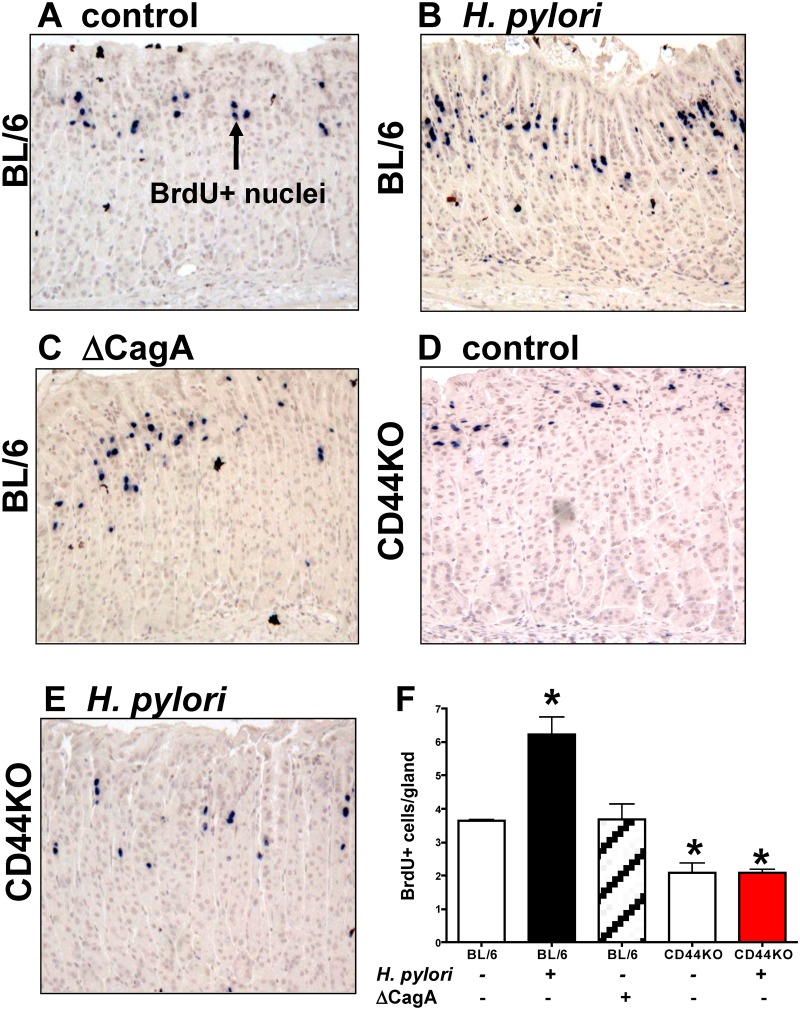
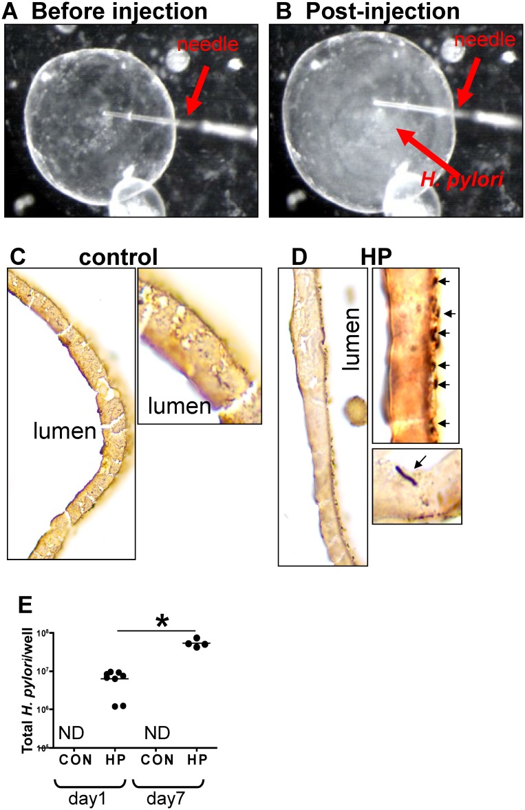
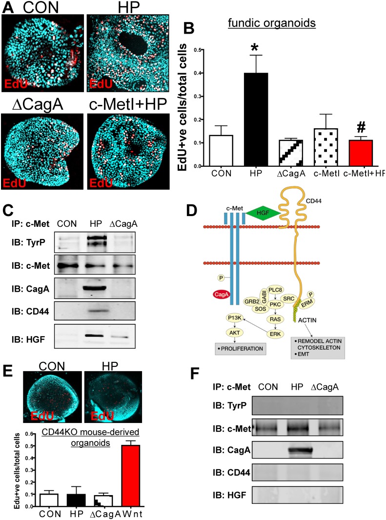
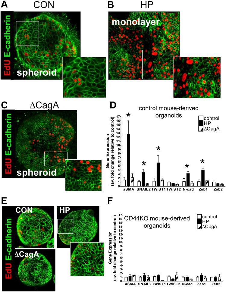
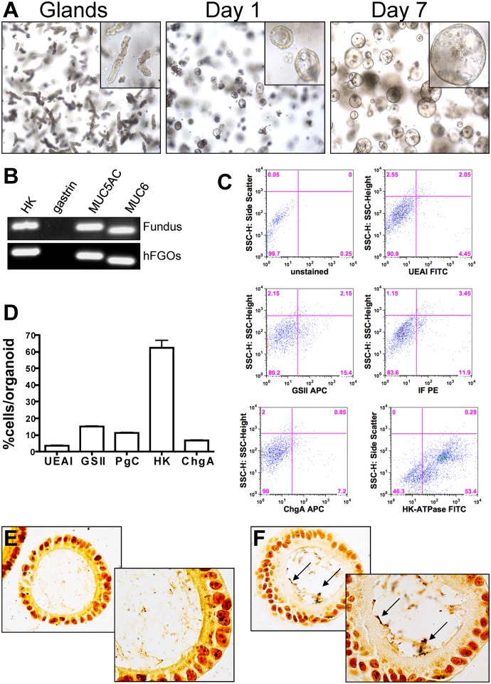
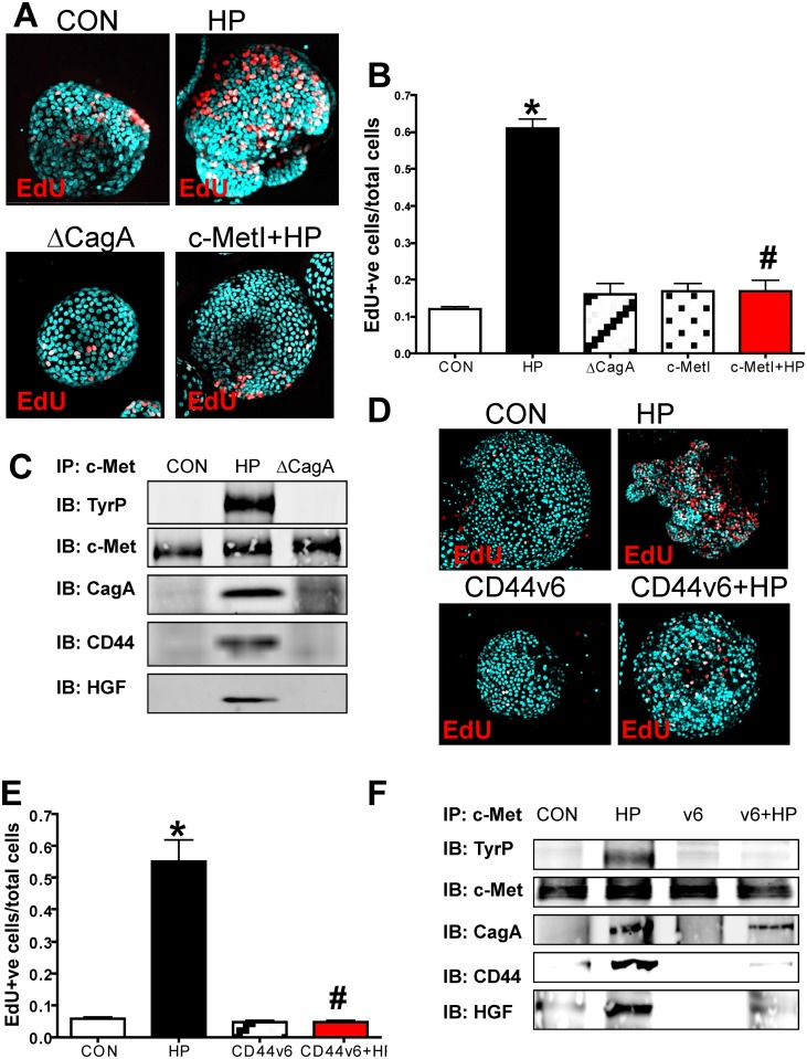
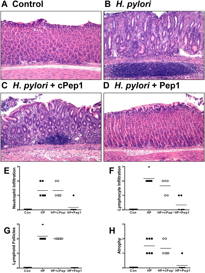
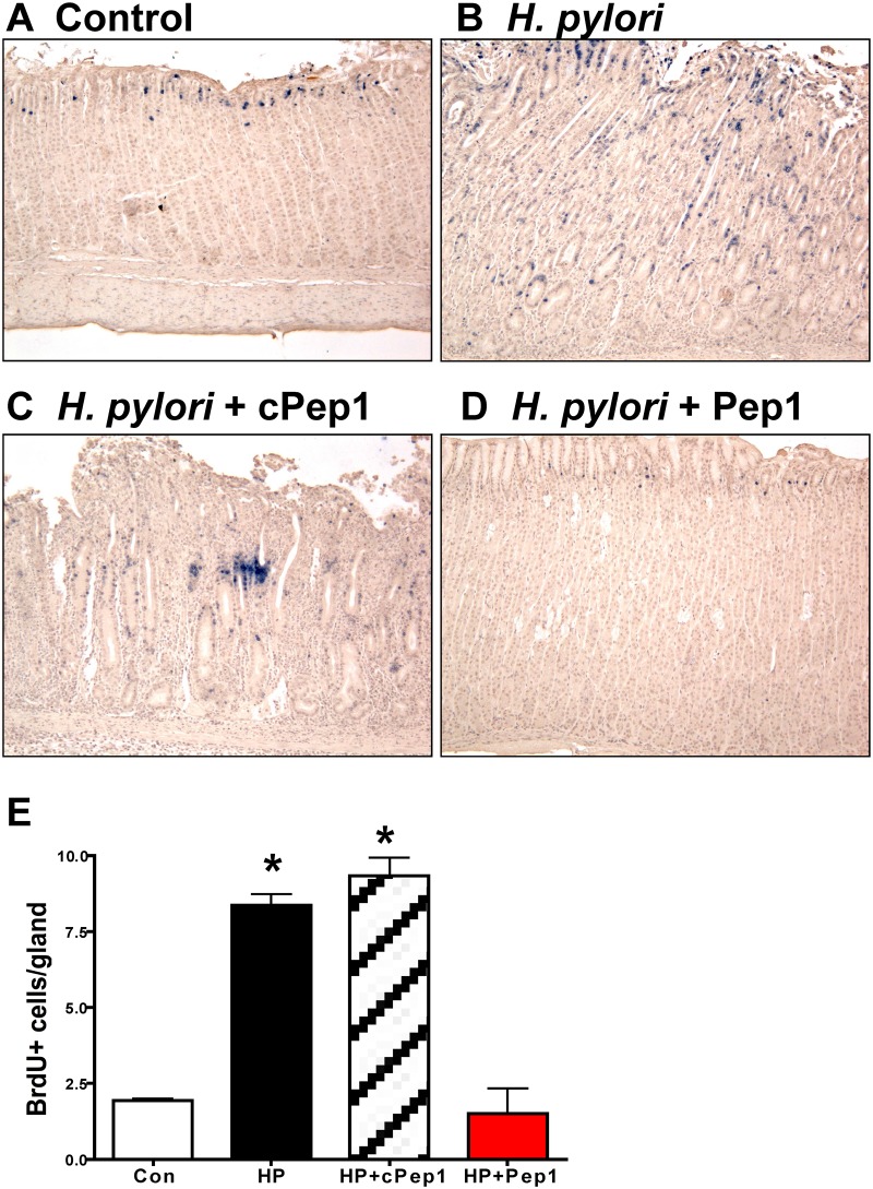
The cytotoxin-associated gene (Cag) pathogenicity island is a strain-specific constituent of Helicobacter pylori (H. pylori) that augments cancer risk. CagA translocates into the cytoplasm where it stimulates cell signaling through the interaction with tyrosine kinase c-Met receptor, leading cellular proliferation. Identified as a potential gastric stem cell marker, cluster-of-differentiation (CD) CD44 also acts as a co-receptor for c-Met, but whether it plays a functional role in H. pylori-induced epithelial proliferation is unknown. We tested the hypothesis that CD44 plays a functional role in H. pylori-induced epithelial cell proliferation. To assay changes in gastric epithelial cell proliferation in relation to the direct interaction with H. pylori, human- and mouse-derived gastric organoids were infected with the G27 H. pylori strain or a mutant G27 strain bearing cagA deletion (∆CagA::cat). Epithelial proliferation was quantified by EdU immunostaining. Phosphorylation of c-Met was analyzed by immunoprecipitation followed by Western blot analysis for expression of CD44 and CagA. H. pylori infection of both mouse- and human-derived gastric organoids induced epithelial proliferation that correlated with c-Met phosphorylation. CagA and CD44 co-immunoprecipitated with phosphorylated c-Met. The formation of this complex did not occur in organoids infected with ∆CagA::cat. Epithelial proliferation in response to H. pylori infection was lost in infected organoids derived from CD44-deficient mouse stomachs. Human-derived fundic gastric organoids exhibited an induction in proliferation when infected with H. pylori that was not seen in organoids pre-treated with a peptide inhibitor specific to CD44. In the well-established Mongolian gerbil model of gastric cancer, animals treated with CD44 peptide inhibitor Pep1, resulted in the inhibition of H. pylori-induced proliferation and associated atrophic gastritis. The current study reports a unique approach to study H. pylori interaction with the human gastric epithelium. Here, we show that CD44 plays a functional role in H. pylori-induced epithelial cell proliferation.


Conflict of interest statement

The authors have declared that no competing interests exist.

Figures

Fig 1
Fig 1. Epithelial proliferation in response to H. pylori infection of C57BL/6 (BL/6) and CD44 deficient (CD44KO) mice.
Stomach sections collected from (A) uninfected BL/6 control, (B) H. pylori infected BL/6, (C) ∆CagA H. pylori strain infected BL/6, and (D) uninfected and (E) H. pylori infected CD44KO mice were immunostained for BrdU incorporation (blue nuclei). (F) Quantification of BrdU+ cells/gland in each group. *P<0.05 compared to controls, n = 6 mice per group.
Fig 2
Fig 2. H. pylori microinjection and colonization of mFGOs.
Organoids were microinjected with H. pylori shown are mFGOs (A) before and (B) after injection. Arrow indicates injection needle. ‘Cloud’ of H. pylori observed after injection is shown. Twenty-four hours after luminal microinjection, H. pylori (HP) attach to the luminal surface demonstrated by Warthin-Starry stain of (C) control and (D) HP infected organoids. (E) Quantitative cultures of HP of control and HP infected organoids. *P<0.05 compared to bacterial numbers at day 1 after infection.
Fig 3
Fig 3. Changes in proliferation in H. pylori infected mFGOs.
(A) Immunofluorescence of EdU+ cells in control (CON), H. pylori (HP) and HP ∆CagA infected and HP infected fundic organoids pretreated with c-Met inhibitor (c-MetI+HP). (B) Quantification of EdU+ cells/total cells. *P<0.05 compared to CON group, n = 6 individual organoid preps per group. (C) Protein lysates were prepared from mFGOs that were uninfected (CON), HP or ∆cagA infected for 24 hours. Lysates were immunoprecipitated using an anti-c-Met antibody and immunoblotted for TyrP, c-Met, CagA, CD44 and HGF. (D) Proposed model of the CD44 co-receptor function in response to H. pylori. (E) Immunofluorescence of EdU+ cells in CON, and HP infected organoids derived from CD44KO mouse fundus. Quantification of EdU+ cells in CON, HP and or ∆CagA infected and Wnt agaonist-treated (Wnt) organoids derived from CD44KO mouse fundus. *P<0.05 compared to CON group, n = 3 individual organoid preps per group. (F) Protein lysates were prepared from mFGOs derived from CD44-deficient mice that were uninfected (CON), HP or ∆CagA infected for 24 hours. Lysates were immunoprecipitated using an anti-c-Met antibody and immunoblotted for TyrP, c-Met, CagA, CD44 and HGF.
Fig 4
Fig 4. H. pylori-induced epithelial-to-mesenchymal transition (EMT).
Immunofluorescence of EdU (red) and E-cadherin (green) using (A) uninfected control (CON), (B) H. pylori (HP) or (C) ∆CagA infected mFGOs. (D) EMT marker (αSMA, SNAIL, TWIST, N-cadherin and Zeb) expression measured by qRT-PCR using RNA isolated from mFGOs uninfected (control), H. pylori (HP) or ∆CagA infected. *P<0.05 compared to CON group, n = 4 individual experiments/group. (E) Immunofluorescence of EdU (red) and E-cadherin (green) using uninfected control (CON), H. pylori (HP) and ∆CagA infected mFGOs derived from stomachs of CD44-deficient mice. (F) (D) EMT marker (αSMA, SNAIL, TWIST, N-cadherin and Zeb) expression measured by qRT-PCR using RNA isolated from mFGOs derived from CD44-deficient mouse stomachs uninfected (control), H. pylori (HP) or ∆CagA infected.
Fig 5
Fig 5. Development and characterization of hFGOs.
(A) Growth of human fundic glands into 3-dimensional epithelial spheres at day 1 and day 7. (B) The hFGOs express markers normally found in human fundus including H+/K+ ATPase (HK), Muc5ac, and Muc6, but lack expression of the antral specific marker gastrin. (C) Flow cytometric analysis of lineage markers for human fundus including UEAI, GSII, pepsinogen C (PgC), Chromogranin A (ChgA), and H+/K+ ATPase (HK). (D) Quantification of flow cytometric data shown in (C). Warthin-Starry staining on (E) uninfected control and (F) H. pylori injected hFGOs.
Fig 6
Fig 6. Changes in proliferation in H. pylori infected hFGOs.
(A) Immunofluorescence of EdU+ cells in control (CON), H. pylori (HP) and HP ΔCagA infected and HP infected fundic organoids pretreated with c-Met inhibitor (c-MetI+HP). (B) Quantification of EdU+ cells/total cells. *P<0.05 compared to CON group, n = 6 individual organoid preps per group. (C) Protein lysates were prepared from hFGOs that were uninfected (CON), HP or ΔcagA infected for 24 hours. Lysates were immunoprecipitated using an anti-c-Met antibody and immunoblotted for TyrP, c-Met, CagA and CD44. (D) Immunofluorescence of EdU+ cells in control (CON), H. pylori (HP) infected, CD44v6 neutralizing antibody treated (CD44v6) and HP infected pretreated with CD44v6 neutralizing antibody (CD44v6+HP) hFGOs. (E) Quantification of EdU+ve cells/total cells. *P<0.05 compared to CON group, #P<0.05 compared to HP infected group, n = 4 individual organoid preps per group. (F) Protein lysates were prepared from hFGOs that were uninfected (CON), H. pylori (HP) infected, CD44v6 neutralizing antibody treated (CD44v6) and HP infected pretreated with CD44v6 neutralizing antibody (CD44v6+HP). Lysates were immunoprecipitated using an anti-c-Met antibody and immunoblotted for TyrP, c-Met, CagA and CD44.
Fig 7
Fig 7. Inhibition of CD44 blocks the development of atrophic gastritis in H. pylori infected Mongolian gerbils.
Mongolian gerbils were administered (A) Brucella broth (control), (B) H. pylori strain 7.13 alone, and in combination with either a (C) control peptide (cPep1), or (D) peptide targeting CD44 (Pep1). Gerbils were sacrificed after 6 weeks infection and H&E’s were scored using the updated Sydney classification for (E) neutrophil infiltration, (F) lymphocyte infiltration, (G) lymphoid follicles, and (H) atrophy.
Fig 8
Fig 8. Epithelial proliferation in response to H. pylori infection in Mongolian gerbils.
Stomach sections collected from Mongolian gerbils administered (A) Brucella broth (control), (B) H. pylori strain 7.13 alone, and in combination with either a (C) control peptide (cPep1), or (D) peptide targeting CD44 (Pep1) were immunostained for BrdU incorporation (blue nuclei). (E) Quantification of BrdU+ cells/gland in each group. *P<0.05 compared to controls, n = 6 gerbils per group.

References

    1. Marshall BJ, Warren JR (1984) Unidentified curved bacilli in the stomach of patients with gastritis and peptic ulceration. Lancet 1: 1311–1315. - PubMed
    1. Correa P, Haenszel W, Cuello C, Tannenbaum S, Archer M (1975) A model for gastric cancer epidemiology. Lancet 2: 58–60. - PubMed
    1. Kuipers EJ, Meuwissen SG (1996) Helicobacter pylori and gastric carcinogenesis. Scand J Gastroenterol 218: 103–105. - PubMed
    1. Kuipers EJ, Perez-Perez GI, Meuwissen SGM, Blaser MJ (1995) Helicobacter pylori and atrophic gastritis: Importance of the cagA status. J Natl Cancer Inst 87: 1777–1780. - PubMed
    1. Kusters JG, van Vliet AH, Kuipers EJ (2006) Pathogenesis of Helicobacter pylori infection. Clin Microbiol Rev 19: 449–490. - PMC - PubMed

Publication types

MeSH terms