. 2015 Feb-Mar;38(2):41-53.
doi: 10.1097/CJI.0000000000000067.

Virological and preclinical characterization of a dendritic cell targeting, integration-deficient lentiviral vector for cancer immunotherapy

Affiliations
Free PMC article

Virological and preclinical characterization of a dendritic cell targeting, integration-deficient lentiviral vector for cancer immunotherapy

Jared M Odegard et al. J Immunother. 2015 Feb-Mar.
Free PMC article

Abstract

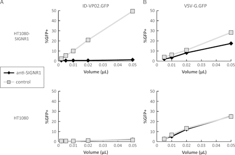
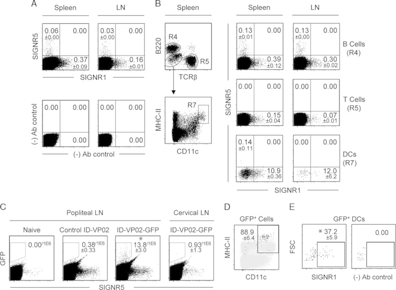
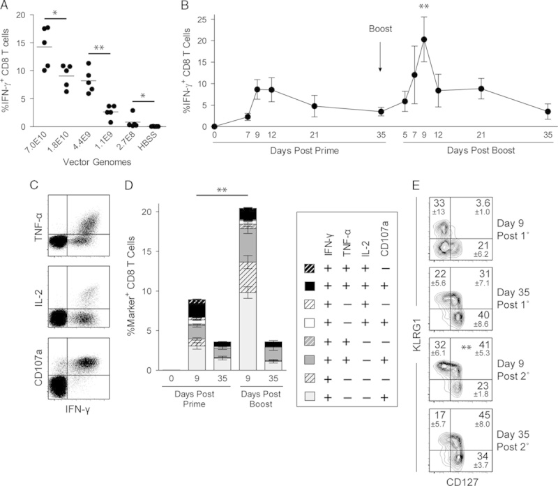
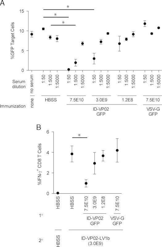
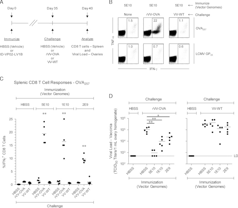
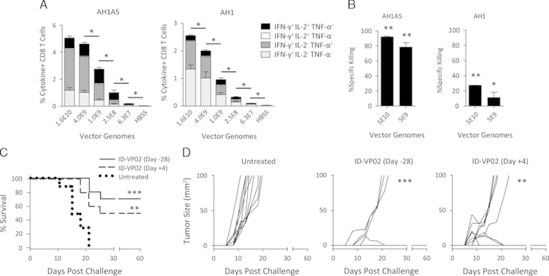
Dendritic cells (DCs) are essential antigen-presenting cells for the initiation of cytotoxic T-cell responses and therefore attractive targets for cancer immunotherapy. We have developed an integration-deficient lentiviral vector termed ID-VP02 that is designed to deliver antigen-encoding nucleic acids selectively to human DCs in vivo. ID-VP02 utilizes a genetically and glycobiologically engineered Sindbis virus glycoprotein to target human DCs through the C-type lectin DC-SIGN (CD209) and also binds to the homologue murine receptor SIGNR1. Specificity of ID-VP02 for antigen-presenting cells in the mouse was confirmed through biodistribution studies showing that following subcutaneous administration, transgene expression was only detectable at the injection site and the draining lymph node. A single immunization with ID-VP02 induced a high level of antigen-specific, polyfunctional effector and memory CD8 T-cell responses that fully protected against vaccinia virus challenge. Upon homologous readministration, ID-VP02 induced a level of high-quality secondary effector and memory cells characterized by stable polyfunctionality and expression of IL-7Rα. Importantly, a single injection of ID-VP02 also induced robust cytotoxic responses against an endogenous rejection antigen of CT26 colon carcinoma cells and conferred both prophylactic and therapeutic antitumor efficacy. ID-VP02 is the first lentiviral vector which combines integration deficiency with DC targeting and is currently being investigated in a phase I trial in cancer patients.


Figures

FIGURE 1
FIGURE 1
Antibody against SIGNR1 blocks transduction with ID-VP02. HT1080 cells expressing SIGNR1 (HT1080-SIGNR1) or the parental cells (HT1080) were preincubated with anti-SIGNR1 antibody (10 µg/mL) for 1 hour, after which increasing doses of (A) ID-VP02 or (B) VSV-G vectors encoding GFP were added. Transduction was assessed 72 hours later. Nevirapine was included on highest vector dose as a control for real transduction (data not shown). Vector titers of ID-VP02 and VSV-G were comparable (115 µg/mL p24, and 111 µg/mL p24, respectively).
FIGURE 2
FIGURE 2
Expression of mouse SIGNR1 and SIGNR5 and ID-VP02 transduction of dendritic cell (DC) in vivo. A, The expression of SIGNR1 and SIGNR5 on spleen and lymph cells was analyzed on live, single-cell events. Control staining pattern lacking SIGNR1-specific and SIGNR5-specific antibodies is shown (fluorescence minus two). B, Live, single-cell events from spleen and lymph node were subdivided into B cells (B220+ TCRβ, labeled R4) and T cells (TCRβ+, B220, labeled R5), and DCs (B220 TCRβ MHC II+ CD11chi, labeled R7) and subsequently analyzed for expression of SIGNR5 and SIGNR1. For all subsets, frequencies of positive events were ≤0.00 in negative control stains lacking SIGNR1-specific and SIGNR5-specific antibodies (data not shown). C, Live, singlet events from the popliteal (draining) or cervical (nondraining) lymph nodes, not gated for any cellular markers, were analyzed for GFP expression. Lymph node cells were pooled from 5 mice, and 3 independent pools were analyzed. Popliteal lymph node cells from naive mice or mice injected with control vector served as negative controls. *P≤0.05 versus non–GFP-encoding control ID-VP02 (Mann-Whitney). D, Frequency of CD11c and MHC II on all GFP+ events, as identified in (C), from the popliteal lymph nodes of mice injected with GFP-encoding ID-VP02 are shown as black dots overlayed on total B220 TCRβ events, shown in gray, as a reference. Gate values are the mean % CD11c+ MHC II+ of the GFP+ cells from 3 independent lymph node pools (5 donors each)±SD. E, Expression of SIGNR1 on GFP+ CD11c+ MHC II+ events, as identified in (D), is shown with (left panel) and without (fluorescence minus one, right panel) inclusion of SIGNR1-specific antibody. *P≤0.05 versus FMO control (Mann-Whitney). Gate statistics are the mean value±SD of 3 biological replicates. Values in (C) are number of positive events per 1×10⁶ cells, whereas all other gate values are percentages.
FIGURE 3
FIGURE 3
ID-VP02 induces high-quality multifunctional primary CD8 T-cell responses that can be effectively expanded with a homologous boost. A, C57BL/6 mice were immunized with indicated doses (vector genomes) of ID-VP02 encoding full-length OVA or HBSS vehicle alone. At day 12 postimmunization, the percentage of OVA257-specific splenic CD8 T cells was measured by intracellular cytokine staining (ICS). B, The kinetics of the primary and secondary CD8 T-cell response to ID-VP02 encoding OVA was determined by immunizing mice (5 per group) with 1×10¹⁰ vector genomes of ID-VP02 in a prime-boost regimen with a 35-day interval and analyzing splenic CD8 T-cell responses at the indicated timepoints. Immunizations were staggered such that all groups were analyzed by ICS on the same day. Peak of the secondary response was significantly greater than the peak of primary response (**P≤0.01, Mann-Whitney). C, Representative intracellular IFN-γ, TNF-α, and IL-2, and surface CD107a staining on viable CD8 T cells after peptide restimulation. D, Frequency of CD8 T cells expressing combinations of IFN-γ, TNF-α, IL-2, and CD107a around the peak and postcontraction of the primary and secondary responses. Negligible numbers of CD8 T cells that were IFN-γ expressed any other effector molecule. E, The effector/memory phenotype of CD44hi H-2Kb-OVA257 pentamer+ CD8 T cells was assessed by staining with CD127 and KLRG1 at the indicated timepoints. **Frequency of CD127+ KLRG1+ cells were significantly greater on day 9 post 2° than on day 9 post 1°. Gate values are mean±SD. *P≤0.05 and **P≤0.01 between indicated groups (Mann-Whitney).
FIGURE 4
FIGURE 4
Neutralizing antibody responses against ID-VP02 can be detected after immunization. A, Ten-fold dilutions of serum taken from mice 28 days after immunization with the indicated doses of ID-VP02 (ID-VP02-GFP), VSV-G pseudotyped vector (VSV-G-GFP), or HBSS, were preincubated with a reporter vector (ID-VP02 encoding GFP) for 1 hour. This serum-vector mix was then used as test article in a GFP transduction assay utilizing 293T.huDC-SIGN target cells. The percentage of GFP+ cells was analyzed 2 days posttransduction. The results are presented as mean±SD of 3 mice per group. B, Groups of mice were first immunized with either 7.5×10¹⁰, 3.0×10⁹, or 1.2×10⁸ vector genomes of ID-VP02 encoding GFP, 7.5×10¹⁰ vector genomes of VSV-G pseudotyped lentivector encoding GFP, or HBSS. On day 28 postprimary immunization the animals were immunized with 3.0×10⁹ of ID-VP02 encoding an alternative antigen cassette, LV1b. OVA257-specific CD8 T-cell response in the spleen was measured by intracellular cytokine staining (ICS) on day 12 after second immunization. *P≤0.05 between indicated groups (Mann-Whitney).
FIGURE 5
FIGURE 5
ID-VP02 immunization induces CD8 T cells that respond and provide protection against viral challenge. A, Experimental schedule. C57BL/6 mice (5 per group) were immunized with 5×10¹⁰, 1×10¹⁰, or 2×10⁹ vector genomes of ID-VP02 encoding a polyepitope antigen (LV1b) that contains the H-2b-restricted OVA257 and LCMV GP33 CD8 T-cell epitopes and then challenged on day 35 postimmunization with 1×10⁷ TCID50 wild-type WR-strain vaccine virus (VV-WT), WR-strain recombinant OVA vaccine virus (rVV-OVA), or left unchallenged. On day 40 (day 5 postchallenge) splenic CD8 T-cell responses and viral load in the ovaries were measured. B, OVA257-specific and LCMV GP33-specific CD8 T-cell responses were measured by staining for intracellular IFN-γ and TNF-α after ex vivo peptide restimulation. Representative dot plots of the CD8 T-cell cytokine profile is shown. C, Frequency of OVA257-specific IFN-γ+ CD8 T cells in each animal. CD8 responses after rVV-OVA challenge were significantly greater in animals immunized ID-VP02 (any dose) compared with vehicle (**P≤0.01). D, Viral load (measured by TCID50 assay) within the ovaries of each animal. *P≤0.05 and **P≤0.01 between indicated groups (Mann-Whitney).
FIGURE 6
FIGURE 6
Prophylactic and therapeutic antitumor efficacy following a single immunization with ID-VP02. A, BALB/c mice (5 per group) were immunized with indicated doses, in vector genomes, of ID-VP02 encoding AH1A5, a heteroclitic mutant of the endogenous CT26 tumor rejection epitope AH1, linked to OVA (OVA-AH1A5) or HBSS vehicle alone. At day 12 postimmunization, the percentage of AH1A5-specific or AH1-specific splenic CD8 T cells was measured by ICS. *P≤0.05 and between indicated groups (Mann-Whitney). B, Twelve days after immunization, a 1:1:1 mixture of dye-labeled target cells each pulsed with AH1, AH1A5, or a control peptide were transferred intravenously into immunized and naive mice (3 per group). The following day, spleens were harvested and the relative recovery of each population was compared between naive and immunized mice to calculate specific killing. *P≤0.05 and **P≤0.01 compared with naive (Mann-Whitney). C and D, BALB/c mice (10 per group) were injected subcutaneously with 8×10⁴ CT26 tumor cells on the right flank and mice were euthanized when tumors exceeded 100 mm2. Mice were either left untreated, treated prophylactically 28 days before challenge with 4×10⁹ vector genomes of ID-VP02 encoding OVA-AH1A5, or treated therapeutically 4 days postchallenge with the same dose of vector. Survival curves are shown in (C) and individual tumor growth curves are shown in (D). **P≤0.01 and ***P≤0.001 compared with untreated (Mantel-Cox).

Similar articles

Cited by

References

    1. Mellman I. Dendritic cells: master regulators of the immune response. Cancer Immunol Res. 2013;1:145–149. - PubMed
    1. Palucka K, Banchereau J. Dendritic-cell-based therapeutic cancer vaccines. Immunity. 2013;39:38–48. - PMC - PubMed
    1. Liu MA. Immunologic basis of vaccine vectors. Immunity. 2010;33:504–515. - PubMed
    1. Yang L, Yang H, Rideout K, et al. Engineered lentivector targeting of dendritic cells for in vivo immunization. Nat Biotechnol. 2008;26:326–334. - PMC - PubMed
    1. Breckpot K, Emeagi PU, Thielemans K. Lentiviral vectors for anti-tumor immunotherapy. Curr Gene Ther. 2008;8:438–448. - PubMed

Publication types

MeSH terms