. 2015 Feb 6;11(2):e1004631.
doi: 10.1371/journal.ppat.1004631. eCollection 2015 Feb.

Cytoplasmic actin is an extracellular insect immune factor which is secreted upon immune challenge and mediates phagocytosis and direct killing of bacteria, and is a Plasmodium Antagonist

Affiliations

Cytoplasmic actin is an extracellular insect immune factor which is secreted upon immune challenge and mediates phagocytosis and direct killing of bacteria, and is a Plasmodium Antagonist

Simone L Sandiford et al. PLoS Pathog. .

Abstract

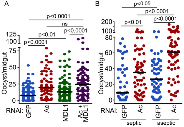
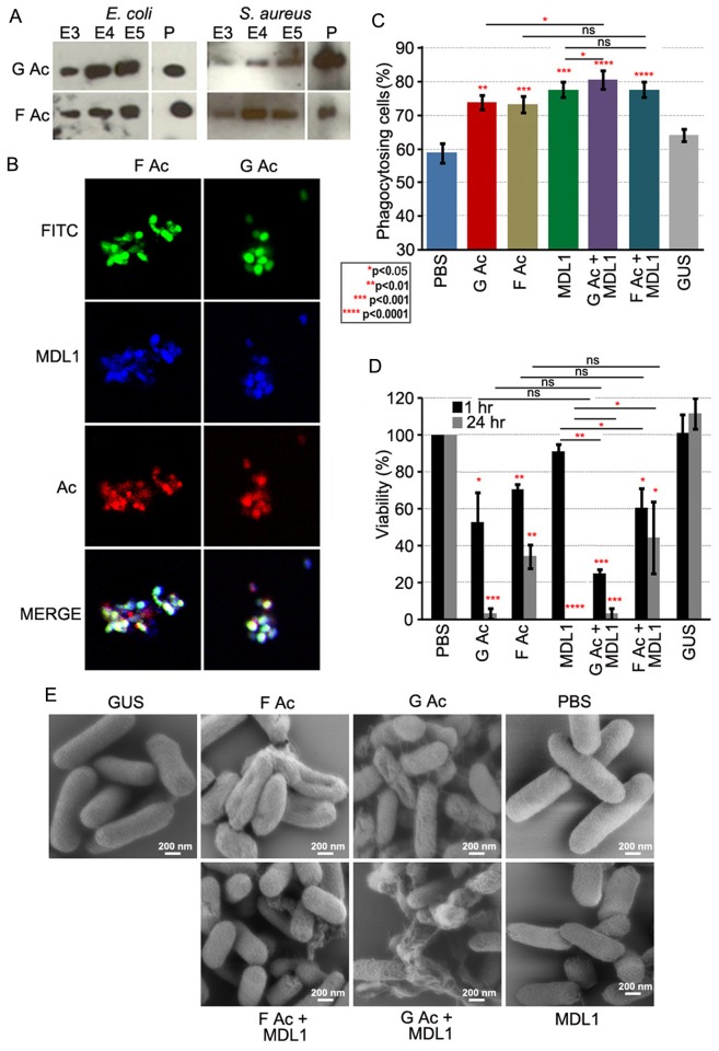
Actin is a highly versatile, abundant, and conserved protein, with functions in a variety of intracellular processes. Here, we describe a novel role for insect cytoplasmic actin as an extracellular pathogen recognition factor that mediates antibacterial defense. Insect actins are secreted from cells upon immune challenge through an exosome-independent pathway. Anopheles gambiae actin interacts with the extracellular MD2-like immune factor AgMDL1, and binds to the surfaces of bacteria, mediating their phagocytosis and direct killing. Globular and filamentous actins display distinct functions as extracellular immune factors, and mosquito actin is a Plasmodium infection antagonist.


Conflict of interest statement

The authors have declared that no competing interests exist.

Figures

Fig 1
Fig 1. An. gambiae actins display tissue, developmental-specific, and immune-responsive expression.
Transcript abundance of all An. gambiae actin genes in the (A) thorax, abdomen, and midgut adult tissues or (B) at distinct developmental stages compared to the adult female mosquito and normalized using the An. gambiae ribosomal S7 gene. Actin 5C (651 A, B, C); larval muscle actin (5095); adult muscle actin (1516); minor actins (1676) and (2127). Expression of actin 5C (651 A, B, C) and adult muscle actin (1516) in An. gambiae Sua5B cells after challenge with (C) LPS (10 μg/mL) (D) Lys-PGN (20 μg/mL) or (E) DAP-PGN (1 μg/mL) for 1,3,6 and 24 hr or (F) live E. coli and S. aureus (MOI 100) for 30 min or 2 hr, compared to non-challenged cells and normalized using the An. gambiae S7 gene.
Fig 2
Fig 2. Actin is secreted into the cell culture supernatant fraction upon immune challenge via an exosome independent mechanism that is regulated by immune pathways.
(A) Immune-challenged (LPS-Pa, LPS-EcK12, LPS-Ec; 10 μg/mL Lys-PGN; 20 μg/mL, or DAP-PGN; 1 μg/mL) Sua5B (An. gambiae), and S2 (D. melongaster) insect cell supernatants and soluble lysate fractions examined for the presence of actin (upper panel) or tubulin (lower panel). (B) Viability of Sua5B and S2 cell lines after treatment with LPS-Pa (10 μg/mL), Lys-PGN (20 μg/mL) or DAP-PGN (1 μg/mL) for 24 hr was determined using the Cell Titer Fluor Cell Viability Assay. (C) Cell viability of Sua5B and S2 cells after saponin (1–10−6%)-induced cell lysis and (D) the amount of actin released into the supernatant was determined for Sua5B (upper panel) and S2 cells (lower panel) at varying saponin concentrations (10−5, 10−4, 10−3 and 1%). (E) Supernatant fractions of Sua5B and Moss55 cells challenged with LPS (10 μg/mL) or Lys-PGN (20 μg/mL) for 24 hr in the presence of 5 μM GW4869 or DMSO (control) and probed for actin. (F) Exosomes (EXO) and lysate fractions isolated from LPS and Lys-PGN stimulated Sua5B or Moss55 cells analyzed for actin. (G) Supernatant fraction of caspar and cactus silenced Sua5B cells probed for the presence of actin.
Fig 3
Fig 3. Actin binds to the surface of different bacteria and is externalized into the mosquito hemolymph upon immune challenge.
Analysis of actin content in the (A) 0.3 M NaCl-eluted fraction of Gram-negative (E. coli, E.c; Esp_Z, E.z.) and Gram-positive (S. aureus, S. a.; B. pumilus B. p.) bacterial species after incubation with immune-challenged cell line supernatants, or (B) 0.3–0.5 M NaCl (E3–E5)-eluted fractions and pellets (P) of Gram-negative (E. coli) and Gram-positive (S. aureus) bacteria incubated with recombinant actin alone (Ac), or together with AgMDL1 (Ac+MDL1). (C) Mosquito hemolymph probed for actin (Ac) and AgMDL1 (MDL1) after challenge with PBS, E. coli (E.c. OD 2.8) or LPS-Pa (100 ng) for 4 hr. (D) Actin content in Moss55 cell supernatant after incubation with E. coli (E.c. MOI 1000) for 30 min, 1 hr, or 2 hr or untreated (UT).
Fig 4
Fig 4. Actin is an extracellular immune factor.
(A) Incubation of E. coli and S. aureus with actin (Ac), AgMDL1 (MDL1), actin +AgMDL1 (Ac+MDL1) or the control protein GUS. Untreated bacteria were incubated with PBS. The percentage of phagocytosing cells was calculated as the number of S2 cells containing at least one phagocytized FITC-labeled bacterium compared to the total number of cells in the field. For each experiment at least 16 fields were counted and the data are representative of three independent experiments. Each bar represents the mean ± the standard deviation. Statistical significance was determined using Student’s t-test.(B) Fluorescent microscopy of S2 cells (red) and FITC-labeled E. coli or S. aureus bacteria (green) incubated with the recombinant proteins actin (Ac) or AgMDL1 (MDL1) alone, or both together (Ac+MDL1) or the control protein GUS. Untreated bacteria were incubated with PBS. (C) Viability of E. coli and S. aureus was determined after 1 or 24 hr of incubation with recombinant proteins (Ac, MDL1, Ac+MDL1, GUS) and compared to untreated bacteria. Error bars represent the mean ± the standard deviation. Statistical significance was determined using Student’s t-test. (D) An. gambiae survival rates after silencing of actin, AgMDL1, actin and AgMDL1, or GFP (control) and challenge of female mosquitoes with E. coli (OD 1.5) or S. aureus (OD 0.4) four days later. Three biological experiments were performed and combined and statistical analysis consisted of a log-rank test to determine the overall significance between all groups, followed by pairwise comparisons between GFP and the other three groups. (E) An. gambiae thorax (6 μg), abdomen (6 μg) and midgut (12 μg) tissues along with hemolymph (2 μg) fractions challenged with LPS (100ng) for 4 hr analyzed for the presence of actin 4 days after silencing of actin or GFP (control). (F) Hemolymph extract from adult female An. gambiae probed for the presence of actin after silencing of GFP (control), actin (Ac) or AgMDL1 (MDL1).
Fig 5
Fig 5. Actin is an antagonist of Plasmodium infection.
P. falciparum oocyst–stage infection intensity after silencing of (A) An. gambiae actin (Ac), AgMDL1 (MDL1), actin and AgMDL1 (Ac+MDL1), or GFP or (B) actin (Ac) or GFP in septic vs aseptic mosquitoes. Circles represents the number of oocysts in an individual mosquito midgut, and the horizontal line indicates the median number. Three independent replicates were obtained, and the Mann-Whitney test was used to determine statistical significance and p-values (indicated above each group).
Fig 6
Fig 6. Globular and filamentous actin display different immune properties.
(A) Actin content in 0.3–0.5M NaCl eluates (E3–E5 fractions) or pellets (P) of E. coli and S. aureus incubated with recombinant globular (G Ac) or filamentous (F Ac) actin. (B) FITC-labeled E. coli (green) incubated with LPS-stimulated Sua5B cell supernatants and stained for AgMDL1 (blue) and actin (red). Co-localization is indicated in white. (C) The percentage of phagocytosing S2 cells containing at least one E. coli bacterium incubated with recombinant globular (G Ac) or filamentous (F Ac) actin with or without AgMDL1 or the control protein GUS as compared to untreated bacteria (PBS). For each assay, at least 16 fields were counted, and the data are representative of three independent experiments. Each bar represents the mean ± the standard deviation. Statistical significance was determined using Student’s t-test. (D) Viability of E. coli was determined after 1- or 24-hr incubations with recombinant globular (G Ac) or filamentous actin (F Ac), with or without AgMDL1, or the control protein GUS as compared to untreated bacteria (PBS). Error bars represent the mean ± the standard deviation. Statistical significance was determined using Student’s t-test. (E) Scanning electron microscopy image of E. coli cells after incubation with G actin (G Ac), F actin (F Ac), AgMDL1 (MDL1) alone or together (G Ac+MDL1, F Ac+MDL1) or with the control protein GUS. Untreated cells were incubated with PBS. Scale bar 200 nm.

Similar articles

Cited by

References

    1. Pollard TD, Cooper JA (2009) Actin, a central player in cell shape and movement. Science 326: 1208–1212. 10.1126/science.1175862 - DOI - PMC - PubMed
    1. Dominguez R, Holmes KC (2011) Actin structure and function. Annu Rev Biophys 40: 169–186. 10.1146/annurev-biophys-042910-155359 - DOI - PMC - PubMed
    1. May RC, Machesky LM (2001) Phagocytosis and the actin cytoskeleton. J Cell Sci 114: 1061–1077. - PubMed
    1. Zhang JG, Czabotar PE, Policheni AN, Caminschi I, Wan SS, et al. (2012) The dendritic cell receptor Clec9A binds damaged cells via exposed actin filaments. Immunity 36: 646–657. 10.1016/j.immuni.2012.03.009 - DOI - PubMed
    1. Ahrens S, Zelenay S, Sancho D, Hanc P, Kjaer S, et al. (2012) F-actin is an evolutionarily conserved damage-associated molecular pattern recognized by DNGR-1, a receptor for dead cells. Immunity 36: 635–645. 10.1016/j.immuni.2012.03.008 - DOI - PubMed

Publication types