. 2015 Feb 6;11(2):e1004669.
doi: 10.1371/journal.ppat.1004669. eCollection 2015 Feb.

Essential domains of Anaplasma phagocytophilum invasins utilized to infect mammalian host cells

Affiliations

Essential domains of Anaplasma phagocytophilum invasins utilized to infect mammalian host cells

David Seidman et al. PLoS Pathog. .

Abstract

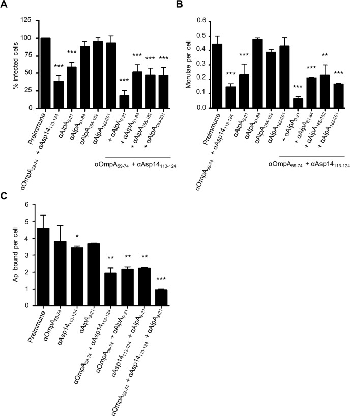
Anaplasma phagocytophilum causes granulocytic anaplasmosis, an emerging disease of humans and domestic animals. The obligate intracellular bacterium uses its invasins OmpA, Asp14, and AipA to infect myeloid and non-phagocytic cells. Identifying the domains of these proteins that mediate binding and entry, and determining the molecular basis of their interactions with host cell receptors would significantly advance understanding of A. phagocytophilum infection. Here, we identified the OmpA binding domain as residues 59 to 74. Polyclonal antibody generated against a peptide spanning OmpA residues 59 to 74 inhibited A. phagocytophilum infection of host cells and binding to its receptor, sialyl Lewis x (sLe(x)-capped P-selectin glycoprotein ligand 1. Molecular docking analyses predicted that OmpA residues G61 and K64 interact with the two sLe(x) sugars that are important for infection, α2,3-sialic acid and α1,3-fucose. Amino acid substitution analyses demonstrated that K64 was necessary, and G61 was contributory, for recombinant OmpA to bind to host cells and competitively inhibit A. phagocytophilum infection. Adherence of OmpA to RF/6A endothelial cells, which express little to no sLe(x) but express the structurally similar glycan, 6-sulfo-sLe(x), required α2,3-sialic acid and α1,3-fucose and was antagonized by 6-sulfo-sLe(x) antibody. Binding and uptake of OmpA-coated latex beads by myeloid cells was sensitive to sialidase, fucosidase, and sLe(x) antibody. The Asp14 binding domain was also defined, as antibody specific for residues 113 to 124 inhibited infection. Because OmpA, Asp14, and AipA each contribute to the infection process, it was rationalized that the most effective blocking approach would target all three. An antibody cocktail targeting the OmpA, Asp14, and AipA binding domains neutralized A. phagocytophilum binding and infection of host cells. This study dissects OmpA-receptor interactions and demonstrates the effectiveness of binding domain-specific antibodies for blocking A. phagocytophilum infection.


Conflict of interest statement

The authors have declared that no competing interests exist.

Figures

Fig 1
Fig 1. OmpA amino acids 59 to 74 are critical for A. phagocytophilum to bind to sLex-capped PSGL-1 and for infection of mammalian host cells.
(A) ELISA in which OmpA23–40, OmpA41–58, and OmpA59–74 antibodies (diluted 1:1600) were used to screen wells coated with GST, GST-OmpA, GST-OmpA19–74, GST-OmpA75–205, or peptides corresponding to OmpA23–40, OmpA41–58, or OmpA59–74. Results shown are the mean ± SD of triplicate samples and are representative of three independent experiments with similar results. (B) Pretreatment of A. phagocytophilum with OmpA59–74 antibody inhibits infection of HL-60 cells in a dose-dependent manner. DC bacteria were incubated with 200 μg/ml of preimmune serum, 200 μg/ml of serum raised against GST-OmpA, or two-fold serially-diluted concentrations of sera raised against OmpA23–40, OmpA41–58, or OmpA59–74 ranging from 0 to 200 μg/ml and then incubated with HL-60 cells. The infection was allowed to proceed for 24 h after which the cells were fixed and examined using immunofluorescence microscopy to quantify the percentage of infected cells. Results shown are relative to host cells that had been incubated with bacteria exposed to preimmune serum and are representative of three experiments with similar results. (C) OmpA59–74 antibody inhibits A. phagocytophilum binding to sLex-capped PSGL-1. DC bacteria were exposed to preimmune serum, antibodies against OmpA, OmpA23–40, OmpA41–58, or OmpA59–74 and then incubated with PSGL-1 CHO cells. Bacteria that were not exposed to antibodies and incubated with PSGL-1 CHO cells or CHO (-) cells were positive and negative controls, respectively, for bacterial binding. The mean numbers ± SD of bound DC organisms per cell were determined using immunofluorescence microscopy. Results shown are the mean ± SD of six combined experiments. Statistically significant (** P < 0.005; ***P < 0.001) values are indicated.
Fig 2
Fig 2. Molecular docking models of A. phagocytophilum OmpA-sLex interactions.
(A) Predicted tertiary structure for A. phagocytophilum OmpA. The orange portion delineates residues 19 to 74. The red portion corresponds to amino acids 59 to 74, and the gray portion corresponds to residues 75 to 205. The dotted line separates the regions encompassed by residues 19 to 74 and 75 to 205. Residues K60, G61, and K64 positions are indicated. (B) Stick representation of the N-terminal PSGL-1 amino acids 61 to 77 (gray) capped with sLex derived from PDB 1g1s. The sLex glycan extends off of threonine 73. sLex linkages and individual sugar residues are denoted. (C) Electrostatic surface map of A. phagocytophilum OmpA, as generated using the PyMol APBS plugin. The left image is oriented as in (A). The right image is rotated 180° around the y-axis. Positive and negative charges are indicated by blue and red, respectively. The dotted line is a demarcation between the regions encompassed by residues 19 to 74 and 75 to 205, which have overall cationic and anionic surface charges, respectively. (D and E) OmpA and sLex interactions predicted by the Autodock Vina algorithm. OmpA is presented as a gray ribbon model, sLex as a multicolor stick model, and hydrogen bonding by dotted lines. OmpA residue K64 is predicted to interact with α2,3-sialic acid (green) of sLex (D and E). Residue G61 is predicted to interact with either α2,3-sialic acid (D) or α1,3-fucose (blue) of sLex (E). Residue K60 is predicted to interact with β1,3-galactose of sLex (E).
Fig 3
Fig 3. G61 and K64 are essential for recombinant OmpA to optimally bind to mammalian host cells and competitively inhibit A. phagocytophilum infection.
GST-OmpA proteins having the CLNHL peptide inserted between OmpA amino acids 67 and 68 or having G61 and/or K64 mutated to alanine are unable to bind to competitively inhibit A. phagocytophilum infection of mammalian host cells. HL-60 cells were incubated with DC organisms in the presence of GST alone, GST-OmpA, GST-OmpA proteins bearing insertions of CLNHL between the indicated residues (A), or GST-OmpA proteins having the indicated amino acids substituted with alanine (B and C) for 1 h. After washing to remove unbound bacteria, host cells were incubated for 24 h and subsequently examined by immunofluorescence microscopy to determine the percentage of infected cells (A and B) or the mean number (± SD) of morulae per cell (C). Results shown in (A), (B), and (C) are the means ± SD for six to twelve combined experiments. The data presented in panel C are the normalized values of six to twelve experiments. Statistically significant (** P < 0.005; ***P < 0.001) values are indicated. (D) Flow cytometric analysis of His-OmpA and His-OmpA proteins bearing alanine substitutions binding to RF/6A cells. Data are representative of two experiments with similar results.
Fig 4
Fig 4. OmpA interacts with α1,3-fucose on mammalian host cell surfaces.
(A to D) RF/6A cells were treated with α1,3/4-fucosidase (A and C), α2,3/6-sialidase (B and D), or vehicle control (- fucosidase and—sialidase, respectively). Glycosidase- and mock-treated cells were incubated with α1,3/6-fucose-specific lectin, AAL; the α2,3-sialic acid-specific lectin, MAL II; His-OmpA; or His-Asp14. (A) The host cells were fixed and screened using immmunofluorescence microscopy (A and B) or flow cytometry (C and D) to detect lectin, His-OmpA, or His-Asp14 binding to host cells. In A and B, green fluorescence corresponds to lectin (AAL or MAL II) or His-tagged protein (OmpA or Aps14) bound at cell surfaces. Host cell nuclei are stained blue by DAPI. (E) AAL and MAL II competitively inhibit His-OmpA binding to mammalian host cells. RF/6A cells were incubated with AAL and MAL II, after which His-OmpA was added. Following the removal of unbound recombinant protein, His-OmpA bound on RF/6A cell surfaces was detected by flow cytometry. Statistically significant (***P < 0.001) values are indicated. Results shown are representative of three experiments with similar results.
Fig 5
Fig 5. OmpA interacts with 6-sulfo sLex on RF/6A endothelial cell surfaces.
(A) Schematic representations of sLex and 6-sulfo sLex. Below each diagram is a statement denoting monoclonal antibodies that recognize each tetrasaccharide. Individual sugar and glycosidic linkages are indicated. (B and C) 6-sulfo sLex is present in high abundance relative to sLex on RF/6A cells. RF/6A cells were screened with sLex-specific antibodies, CSLEX1 and KM93; 6-sulfo sLex-specific antibody, G72; or IgM isotype control followed by detection of cell surface bound antibodies using immunofluorescence microscopy (B) and flow cytometry (C). (D) Antibody blocking of 6-sulfo sLex inhibits His-OmpA binding to RF/6A cells. RF/6A cells were incubated with CSLEX1, KM93, G72, IgM, or vehicle (Cells only) followed by the addition of His-OmpA, and washing to remove unbound recombinant protein. Flow cytometry was used to detect bound His-OmpA. Statistically significant (***P < 0.001) values are indicated. Results shown are representative of two experiments with similar results.
Fig 6
Fig 6. OmpA coated beads bind to and are internalized by non-phagocytic endothelial cells.
(A) Confirmation of recombinant OmpA conjugation to inert beads. Red fluorescent OmpA-coated microspheres (OmpA beads) were incubated with OmpA antibody (αOmpA) or isotype control. Unconjugated (Control) beads were included as a negative control. Bound antibody (green) was detected using immunofluorescence microscopy. OmpA coated beads with bound antibody appeared yellow when the individual fluorescence channels were merged, whereas beads without bound antibody appeared red. (B to D) OmpA coated beads bind to and are internalized by non-phagocytic RF/6A cells. OmpA coated beads were incubated with RF/6A cells for 1 h after which unbound beads were washed off. Cells were screened with OmpA antibody and examined using immunofluorescence (B and D) or scanning electron microscopy (C) to assess binding or were incubated further to allow for bead uptake. To assess for internalized beads, the host cells were treated with trypsin, washed, incubated overnight for the endothelial cells to re-adhere, fixed, and screened with OmpA antibody and immunofluorescence microscopy. Because the host cells were not permeabilized, bound beads detected by OmpA antibody appeared yellow and internalized beads, which OmpA antibody could not detect, remained red when viewed by immunofluorescence microscopy. (B) DIC, differential interference contrast microscopy. (C) Two scanning electron micrographs depicting bound or internalized OmpA coated beads. Arrows denote filopodia-like structures bound to beads. Scale bars are indicated. Results in (D) are representative of thirteen experiments with similar results. Statistically significant (** P < 0.005; ****P < 0.0001) values are indicated.
Fig 7
Fig 7. OmpA coated bead binding to and uptake by promyelocytic HL-60 cells involve OmpA residues G61 and K64 and are dependent on sLex.
(A) Scanning electron micrographs depicting OmpA coated beads bound to and being internalized by HL-60 cells. Arrows point to filopodia-like structures adhered to beads. Scale bars are indicated. (B and C) HL-60 cells were incubated with beads coated with OmpA, OmpA proteins having the indicated amino acids substituted with alanine, or non-coated control beads. The numbers of bound and internalized beads were determined using immunofluorescence microscopy. (D to I) HL-60 cells were incubated with α2,3/6-sialidase, α1,3/4-fucosidase, or vehicle only (D and E), sLex antibody CSLEX1 or IgM isotype control (F and G), or PSGL-1 N-terminus antibody KPL-1 or IgG isotype control (H and I) before being incubated with OmpA coated or non-coated control beads. The numbers of bound and internalized beads were assessed using immunofluorescence microscopy. Results in (B) through (I) are the mean ± SD of triplicate samples and are representative of three independent experiments with similar results. Statistically significant (** P < 0.005; ***P < 0.001) values are indicated.
Fig 8
Fig 8. The Asp14 binding domain is contained within amino acids 113 to 124.
(A) Pretreatment of A. phagocytophilum with Asp14113–124 antiserum inhibits infection of HL-60 cells in a dose-dependent manner. DC bacteria were incubated with 200 μg/ml of preimmune serum, 200 μg/ml of serum raised against full-length Asp14, or two-fold serially-diluted concentrations of anti-Asp1498–112 or anti-Asp14113–124 ranging from 0 to 200 μg/ml and then incubated with HL-60 cells. The infection was allowed to proceed for 24 h prior to being assessed by immunofluorescence microscopy for the percentage of infected cells. (B) A combination of antisera targeting OmpA59–74 and Asp14113–124 inhibits A. phagocytophilum infection of HL-60 cells better than serum targeting either binding domain alone. DC organisms were exposed to preimmune serum or antisera targeting OmpA59–74, Asp14113–124, OmpA59–74 plus Asp1498–112, or anti-Asp14113–124 together with OmpA59–74, OmpA23–40, or OmpA43–58 antibodies. The cells were fixed and screened using immunofluorescence microscopy to determine the percentages of infected cells. (C and D) OmpA59–74 and Asp14113–124 Fab fragments effectively inhibit A. phagocytophilum infection of HL-60 cells. DC bacteria were incubated with Fab fragments derived from preimmune serum, antibodies targeting OmpA23–40, OmpA41–58, OmpA59–74, Asp1498–112, Asp14113–124, or OmpA59–74 Fab fragment together with Asp14113–124 Fab fragment. The cells were fixed and screened to determine the percentages of infected cells (C) and morulae per cell (D). Results presented in (B) to (D) are relative to host cells that had been incubated with bacteria treated with preimmune serum. Results presented in (A) and (B) are the means ± SD for three experiments. Results in (C) and (D) are the mean ± SD of triplicate samples and are representative of two experiments with similar results. Statistically significant (* P < 0.05; ** P < 0.005; ***P < 0.001) values are indicated.
Fig 9
Fig 9. A combination of antisera targeting the binding domains of OmpA, Asp14, and AipA blocks A. phagocytophilum infection of mammalian host cells.
DC organisms were incubated with preimmune serum or antibodies specific for OmpA59–74 and Asp14113–124; or AipA9–21, AipA61–84, AipA165–182, or AipA183–201, either independently or in combination with OmpA59–74 and Asp14113–124 antibodies. Next, the bacteria were incubated with HL-60 cells. The infection was allowed to proceed for 24 h, after which the host cells were fixed and examined using immunofluorescence microscopy to determine the percentages of infected cells (A) and the number of morulae per cell (B). (C) To verify that the observed reductions in A. phagocytophilum infection were due to antisera mediated blocking of bacterial binding to HL-60 cell surfaces, the experiment was repeated except that DC organisms were incubated with antibodies targeting OmpA59–74 and/or Asp14113–124, and/or AipA9–21 prior to being incubated with host cells, and the numbers of bound bacteria per cell was assessed. Results presented are relative to host cells that had been incubated with bacteria treated with preimmune serum and are the means ± SD for six combined experiments. Statistically significant (* P < 0.05; ** P < 0.005; ***P < 0.001) values are indicated.

References

    1. Truchan HK, Seidman D, Carlyon JA (2013) Breaking in and grabbing a meal: Anaplasma phagocytophilum cellular invasion, nutrient acquisition, and promising tools for their study. Microbes Infect. - PMC - PubMed
    1. CDC (2013) Notice to readers: final 2012 reports of nationally notifiable infectious diseases. MMWR Morb Mortal Wkly Rep 62: 669–682. - PMC - PubMed
    1. Hopkins RS, Jajosky RA, Hall PA, Adams DA, Connor FJ, et al. (2005) Summary of notifiable diseases—United States, 2003. MMWR Morb Mortal Wkly Rep 52: 1–85. - PubMed
    1. Hao Q, Geng Z, Hou XX, Tian Z, Yang XJ, et al. (2013) Seroepidemiological investigation of lyme disease and human granulocytic anaplasmosis among people living in forest areas of eight provinces in China. Biomed Environ Sci 26: 185–189. 10.3967/0895-3988.2013.03.005 - DOI - PubMed
    1. Zhang XC, Zhang LX, Li WH, Wang SW, Sun YL, et al. (2012) Ehrlichiosis and zoonotic anaplasmosis in suburban areas of Beijing, China. Vector Borne Zoonotic Dis 12: 932–937. 10.1089/vbz.2012.0961 - DOI - PubMed

Publication types

MeSH terms

Substances