Dorsiflexion, plantar-flexion, and neutral ankle positions during passive resistance assessments of the posterior hip and thigh muscles
- PMID: 25658906
- PMCID: PMC4560017
- DOI: 10.4085/1062-6050-49.6.04
Dorsiflexion, plantar-flexion, and neutral ankle positions during passive resistance assessments of the posterior hip and thigh muscles
Abstract
Context: Passive straight-legged-raise (SLR) assessments have been performed with the ankle fixed in dorsiflexion (DF), plantar-flexion (PF), or neutral (NTRL) position. However, it is unclear whether ankle position contributes to differences in the passive resistance measured during an SLR assessment.
Objective: To examine the influence of ankle position during an SLR on the passive torque, range of motion (ROM), and hamstrings electromyographic (EMG) responses to passive stretch of the posterior hip and thigh muscles.
Design: Crossover study.
Setting: Research laboratory.
Patients or other participants: A total of 13 healthy volunteers (5 men: age = 24 ± 3 years, height = 178 ± 6 cm, mass = 85 ± 10 kg; 8 women: age = 21 ± 1 years, height = 163 ± 8 cm, mass = 60 ± 6 kg).
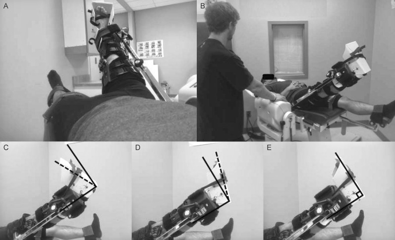
Intervention(s): Participants performed 6 randomly ordered passive SLR assessments involving 2 assessments at each condition, which included the ankle positioned in DF, PF, and NTRL. All SLRs were performed using an isokinetic dynamometer programmed in passive mode to move the limb toward the head at 5°/s.
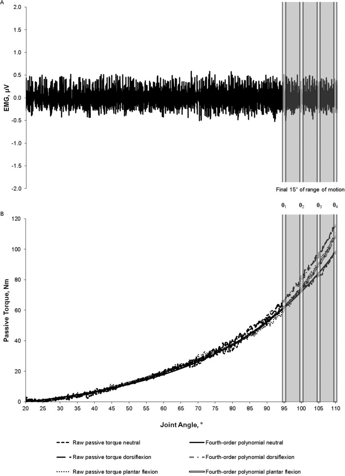
Main outcome measure(s): During each SLR, maximal ROM was determined as the point of discomfort but not pain, as indicated by the participant. Passive torque and EMG amplitude were determined at 4 common joint angles (θ) separated by 5° during the final common 15° of ROM for each participant.
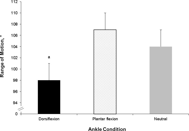
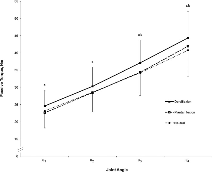
Results: Passive torque was greater for the DF condition than the NTRL (P = .008) and PF (P = .03) conditions at θ3 and greater for the DF than NTRL condition (P = .02) at θ4. Maximal ROM was lower for the DF condition than the NTRL (P = .003) and PF (P < .001) conditions. However, we found no differences among conditions for EMG amplitude (P = .86).
Conclusions: These findings suggest that performing SLRs with the ankle positioned in DF may elicit greater passive torque and lower ROM than SLRs with the ankle positioned in PF or NTRL. The greater passive torque and lower ROM induced by the DF condition possibly were due to increased tension in the neural structures of the proximal thigh.
Keywords: hamstrings muscles; neural tension; passive torque; range of motion; stiffness.
Figures
Similar articles
-
Reliability of manual versus automated techniques for assessing passive stiffness of the posterior muscles of the hip and thigh.J Sports Sci. 2013;31(8):867-77. doi: 10.1080/02640414.2012.753159. Epub 2012 Dec 21. J Sports Sci. 2013. PMID: 23256778
-
Influence of stretching velocity on musculotendinous stiffness of the hamstrings during passive straight-leg raise assessments.Musculoskelet Sci Pract. 2017 Aug;30:80-85. doi: 10.1016/j.msksp.2016.12.018. Epub 2017 Jan 5. Musculoskelet Sci Pract. 2017. PMID: 28715304
-
Effects of hip and head position on ankle range of motion, ankle passive torque, and passive gastrocnemius tension.Scand J Med Sci Sports. 2016 Jan;26(1):41-7. doi: 10.1111/sms.12406. Epub 2015 Feb 12. Scand J Med Sci Sports. 2016. PMID: 25676048
-
Passive properties of human skeletal muscle during stretch maneuvers. A review.Scand J Med Sci Sports. 1998 Apr;8(2):65-77. doi: 10.1111/j.1600-0838.1998.tb00171.x. Scand J Med Sci Sports. 1998. PMID: 9564710 Review.
-
Differences in ankle joint complex range of motion as a function of age.Foot Ankle. 1993 May;14(4):215-22. doi: 10.1177/107110079301400407. Foot Ankle. 1993. PMID: 8359768 Review.
Cited by
-
Safety of lower extremity neurodynamic exercises in adults with diabetes mellitus: a feasibility study.J Man Manip Ther. 2017 Feb;25(1):30-38. doi: 10.1080/10669817.2016.1180772. Epub 2016 Jun 17. J Man Manip Ther. 2017. PMID: 28855790 Free PMC article.
-
Involuntary hamstring muscle activity reduces passive hip range of motion during the straight leg raise test: a stimulation study in healthy people.BMC Musculoskelet Disord. 2019 Mar 27;20(1):130. doi: 10.1186/s12891-019-2511-6. BMC Musculoskelet Disord. 2019. PMID: 30917805 Free PMC article.
-
Remote effects of a 7-week combined stretching and foam rolling training intervention of the plantar foot sole on the function and structure of the triceps surae.Eur J Appl Physiol. 2023 Aug;123(8):1645-1653. doi: 10.1007/s00421-023-05185-5. Epub 2023 Mar 27. Eur J Appl Physiol. 2023. PMID: 36973555 Free PMC article.
-
Myofascial Treatment Techniques on the Plantar Surface Influence Functional Performance in the Dorsal Kinetic Chain.J Sports Sci Med. 2022 Feb 15;21(1):13-22. doi: 10.52082/jssm.2022.13. eCollection 2022 Mar. J Sports Sci Med. 2022. PMID: 35250329 Free PMC article. Clinical Trial.
-
Predictors of dysfunction and health-related quality of life in the flexion pattern subgroup of patients with chronic lower back pain: The STROBE study.Medicine (Baltimore). 2018 Jul;97(29):e11363. doi: 10.1097/MD.0000000000011363. Medicine (Baltimore). 2018. PMID: 30024508 Free PMC article.
References
-
- Ryan ED, Thompson BJ, Herda TJ, et al. The relationship between passive stiffness and evoked twitch properties: the influence of muscle CSA normalization. Physiol Meas. 2011;32(6):677–686. - PubMed
-
- Gajdosik RL. Passive extensibility of skeletal muscle: review of the literature with clinical implications. Clin Biomech (Bristol, Avon) 2001;16(2):87–101. - PubMed
-
- Marshall PWM, Mannion J, Murphy BA. Extensibility of the hamstrings is best explained by mechanical components of muscle contraction, not behavioral measures in individuals with chronic low back pain. PMR. 2009;1(8):709–718. - PubMed
-
- Palmer TB, Jenkins NDM, Thompson BJ, Smith DB, Cramer JT. The relationship between passive stiffness and muscle power output: influence of muscle CSA normalization. Muscle Nerve. 2014;49(1):69–75. - PubMed
-
- Ekstrand J, Gillquist J. The frequency of muscle tightness and injuries in soccer players. Am J Sports Med. 1982;10(2):75–78. - PubMed
MeSH terms
LinkOut - more resources
Full Text Sources
Other Literature Sources
Miscellaneous