. 2015 Feb 6;11(2):e1004938.
doi: 10.1371/journal.pgen.1004938. eCollection 2015 Feb.

Asymmetry of the budding yeast Tem1 GTPase at spindle poles is required for spindle positioning but not for mitotic exit

Affiliations

Asymmetry of the budding yeast Tem1 GTPase at spindle poles is required for spindle positioning but not for mitotic exit

Ilaria Scarfone et al. PLoS Genet. .

Abstract

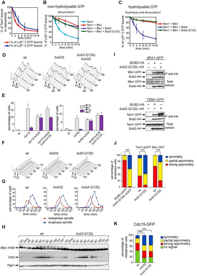
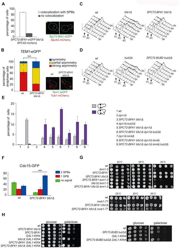
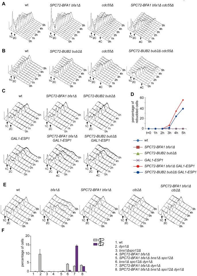
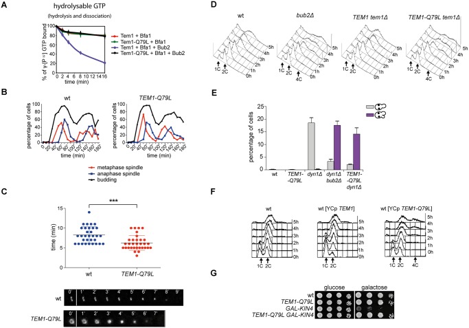
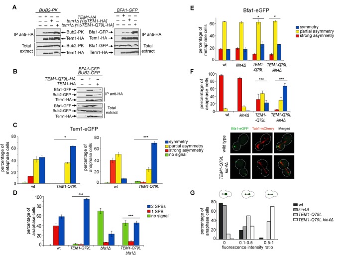
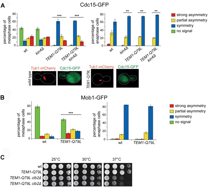
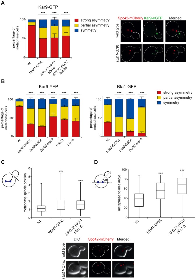
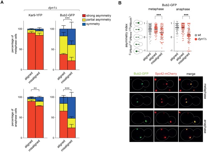
The asymmetrically dividing yeast S. cerevisiae assembles a bipolar spindle well after establishing the future site of cell division (i.e., the bud neck) and the division axis (i.e., the mother-bud axis). A surveillance mechanism called spindle position checkpoint (SPOC) delays mitotic exit and cytokinesis until the spindle is properly positioned relative to the mother-bud axis, thereby ensuring the correct ploidy of the progeny. SPOC relies on the heterodimeric GTPase-activating protein Bub2/Bfa1 that inhibits the small GTPase Tem1, in turn essential for activating the mitotic exit network (MEN) kinase cascade and cytokinesis. The Bub2/Bfa1 GAP and the Tem1 GTPase form a complex at spindle poles that undergoes a remarkable asymmetry during mitosis when the spindle is properly positioned, with the complex accumulating on the bud-directed old spindle pole. In contrast, the complex remains symmetrically localized on both poles of misaligned spindles. The mechanism driving asymmetry of Bub2/Bfa1/Tem1 in mitosis is unclear. Furthermore, whether asymmetry is involved in timely mitotic exit is controversial. We investigated the mechanism by which the GAP Bub2/Bfa1 controls GTP hydrolysis on Tem1 and generated a series of mutants leading to constitutive Tem1 activation. These mutants are SPOC-defective and invariably lead to symmetrical localization of Bub2/Bfa1/Tem1 at spindle poles, indicating that GTP hydrolysis is essential for asymmetry. Constitutive tethering of Bub2 or Bfa1 to both spindle poles impairs SPOC response but does not impair mitotic exit. Rather, it facilitates mitotic exit of MEN mutants, likely by increasing the residence time of Tem1 at spindle poles where it gets active. Surprisingly, all mutant or chimeric proteins leading to symmetrical localization of Bub2/Bfa1/Tem1 lead to increased symmetry at spindle poles of the Kar9 protein that mediates spindle positioning and cause spindle misalignment. Thus, asymmetry of the Bub2/Bfa1/Tem1 complex is crucial to control Kar9 distribution and spindle positioning during mitosis.


Conflict of interest statement

The authors have declared that no competing interests exist.

Figures

Fig 1
Fig 1. Bub2 GAP activity involves a ‘dual finger’ mechanism and promotes Bub2/Bfa1 clearance from the mother SPB.
A-C: Bacterially purified GST-Bub2 or GST-Bub2-Q132L, MBP-Bfa1 and 6xHis-Tem1 proteins were used to measure the kinetics of hydrolysis+dissociation (γ[32P]GTP) or dissociation only (γ[35S]GTP) using a filter binding assay (see Materials and Methods). Graphs show average values and standard deviations from three independent experiments. D: Exponentially growing cultures of the indicated strains were shifted to nocodazole containing medium at t = 0. Cell samples were withdrawn at the indicated time for FACS analysis of DNA contents. E: The percentage of cells with binucleate cell bodies accompanied or not by a checkpoint defect (indicated by re-budding in the absence of proper chromosome segregation) was scored in cycling cultures of the indicated strains shifted either to 14°C for 16h (left graph) or to 37°C for 3h (right graph). F-G: Exponentially growing cells with the indicated genotypes were arrested in G1 by α-factor and released into fresh medium at time 0. At 70’ after release α-factor was re-added to prevent cells from entering a second cell cycle. Cell samples were collected for FACS analysis of DNA contents (F) and for tubulin staining by indirect immunofluorescence (G). H: Cells were treated as in (F-G). TCA extracts were prepared from cell samples at the indicated time points to monitor kinetics of Bfa1-HA6 phosphorylation and Clb2 accumulation and degradation by western blot analysis. Pgk1 was used as loading control. I: Protein extracts from cells expressing the indicated tagged proteins were used for immunoprecipitation with an anti-HA affinity resin. Western blot analysis was then performed with anti-GFP and anti-HA antibodies. The input represents 1/25th of the total extract used for each IP. J-K: Localization of eGFP- tagged Bub2/Bub2-Q132L, Tem1, Bfa1 (J) and Cdc15-GFP (K) was analysed by fluorescence microscopy after formaldehyde fixation.
Fig 2
Fig 2. Constitutive targeting of Bfa1 to SPBs facilitates mitotic exit by recruiting Tem1 to SPBs.
A-B: Cycling cells co-expressing Spc72-Bfa1-eGFPand Spc42-mCherry to mark the SPB (upper panel) or co-expressing Tem1-eGFP and Tub1-GFP (to mark microtubules, lower panel) were analysed to study the distribution of Spc72-Bfa1-eGFP (A) and Tem1-eGFP (B) at SPBs in SPC72-BFA1 bfa1Δ cells. C-D: Cycling cells with the indicated genotypes were shifted into nocodazole containing medium (t = 0). Cell samples were withdrawn at the indicated times for FACS analysis of DNA contents. E: The percentage of cells with binucleate cell bodies accompanied or not by a SPOC defect was scored after propidium iodide staining of cycling cultures of cells with the indicated genotypes after shift to 14°C for 16h. The histograms on the right side represent the DNA contents of the same cells as measured by FACS analysis. F: Percentage of metaphase cells with Cdc15-GFP at 0, 1 or 2 SPBs was scored in the indicated strains after formaldehyde fixation. Metaphases were identified by means of the Tub1-mCherry co-expressed marker. G: Serial dilutions of stationary phase cultures of the indicated strains were spotted on YPD and incubated at the indicated temperature. H: Serial dilutions of stationary phase cultures of the indicated strains were spotted on YP medium containing either glucose or galactose and incubated at 25°C for 48h.
Fig 3
Fig 3. The activation state of the FEAR pathway influences the ability of Spc72-Bfa1 to promote unscheduled mitotic exit.
A-E: Logarithmically growing cultures of cells with the indicated genotypes were synchronized in G1 by α-factor and then released into nocodazole-containing medium (t = 0). At the indicated times cell samples were withdrawn for FACS analysis of DNA contents (A-C, E) and to score the percentage of re-budded cells (D). F: The percentage of cells with binucleate cell bodies accompanied or not by a SPOC defect was scored after DAPI staining of cycling cells with the indicated genotypes shifted to14°C for 16h.
Fig 4
Fig 4. The constitutively active Tem1-Q79L variant is checkpoint-deficient.
A: Bacterially purified 6XHis-Tem1 and 6XHis-Tem1-Q79L were loaded with γ[32P]GTP either in the absence or in the presence of recombinant MBP-Bfa1 and incubated at 30°C for 10 minutes. The mixture was then added to GST-Bub2 or buffer alone and kinetics of GTP hydrolysis and dissociation was followed by a filter-binding assay (see details in Material and Methods). Graphs show average values and standard deviations from three independent experiments. B: Wild type and TEM1-Q79L cells were arrested in G1 by α-factor and then released into fresh medium at 25°C (t = 0). Cell samples were withdrawn every 10’ to measure kinetics of budding and spindle formation/elongation after in situ immunostaining of tubulin. C: Actomyosin ring contraction has been visualized by live cell imaging of wild type and TEM1-Q79L expressing Myo1-GFP (n = 30). D: Logarithmically growing cultures of cells with the indicated genotypes were shifted into nocodazole containing medium (t = 0). DNA contents were analysed by flow cytometry at the indicated times. E: The percentage of cells with binucleate cell bodies accompanied or not by SPOC defect was scored after DAPI staining of cycling cells of the indicated strains shifted to 14°C for 16h. F: Logarithmically growing cultures of strains with the indicated genotypes were shifted to nocodazole containing medium (t = 0). DNA contents were analysed by flow cytometry at the indicated times. G: Serial dilutions of stationary phase cultures of the indicated strains were spotted on YPD or YP galactose plates and incubated at 30°C for 48h.
Fig 5
Fig 5. Constitutively active Tem1-Q79L binds more avidly Bub2/Bfa1 and shows reduced asymmetry at anaphase spindle poles.
A-B: Protein extracts from cells expressing the indicated tagged proteins were used for immunoprecipitation with an anti-HA affinity resin. Western blot analysis was then performed with anti-PK, anti-GFP and anti-HA antibodies. The input represents 1/25th of the total extract used for each IP. C-F: Localization of eGFP- tagged Tem1 and Tem1-Q79L (C-D) or Bfa1-eGFP (E-F) was analysed in the indicated strains by fluorescence microscopy after formaldehyde fixation. Metaphase and anaphase cells were identified by means of the Tub1-mCherry co-expressed marker. Micrographs show representative cells of each strain in anaphase. G: Fluorescence intensity ratios were calculated between the two SPBs in anaphase cells of the indicated strains (see details in Materials and Methods).
Fig 6
Fig 6. The TEM1-Q79L allele leads to premature Cdc15, but not Mob1, loading on SPBs.
A-B: Distribution of Cdc15-GFP (A) or Mob1-GFP (B) was analysed in the indicated strains by fluorescence microscopy after formaldehyde fixation. Metaphase and anaphase cells were identified by means of the Tub1-mCherry co-expressed marker. Micrographs show representative wild type and TEM1-Q79L cells expressing Cdc15-GFP in metaphase. C: Serial dilutions of stationary phase cells with the indicated genotypes were spotted on YPD and incubated at the indicated temperatures for 48h.
Fig 7
Fig 7. Bub2/Bfa1 and Tem1 asymmetry is important for proper Kar9 distribution and spindle positioning.
A-B: Percentage of metaphase cells carrying Kar9-eGFP (A), Kar9-YFP or Bfa1-eGFP (B) on one SPB (strong asymmetry), both SPBs unequally (partial asymmetry) or both SPBs equally (symmetry) for the indicated strains after formaldehyde fixation. Metaphase and anaphase cells were identified by means of the co-expressed SPB marker Spc42-mCherry. C-D: Spindle position (distance between the nearest SPB and the bud neck) and orientation (angle that the spindle forms with respect to the cell polarity axis) were measured in metaphase spindles of cells with the indicated genotypes after formaldehyde fixation (n≥100).
Fig 8
Fig 8. Bub2, but not Kar9, become symmetric on misaligned spindles.
A: Distribution of Kar9-YFP and Bub2-GFP in metaphase and anaphase on aligned and misaligned (dyn1Δ) spindles. Metaphase and anaphase cells were identified by means of the co-expressed SPB marker Spc42-mCherry. Misaligned spindles in metaphase cells were identified by measuring an angle of 60°-120° between the spindle axis and a line connecting the old SPB (defined as the SPB showing brighter Spc42-mCherry fluorescence) to the bud neck. Scoring was done on 3 independent clones for each genotype with a total N ≥ 75; mean ± SD. B: Asymmetry indices were calculated for wild type and dyn1Δ cells expressing Bub2-GFP and Spc42-mCherry in (A) by dividing the difference between Bub2 fluorescence associated with the old SPB (ISPBold) and new SPB (ISPBnew) by the total Bub2 fluorescence (ISPBboth). Age of SPBs was determined by the fluorescence intensity of Spc42-mCherry (bright fluorescence: old SPB, dim fluorescence: new SPB). Micrographs show representative Bub2-GFP distribution on aligned and misaligned spindles, in either metaphase or anaphase.

Similar articles

Cited by

References

    1. Inaba M, Yamashita YM (2012) Asymmetric stem cell division: precision for robustness. Cell Stem Cell 11: 461–469. 10.1016/j.stem.2012.09.003 - DOI - PubMed
    1. Knoblich JA (2010) Asymmetric cell division: recent developments and their implications for tumour biology. Nat Rev Mol Cell Biol 11: 849–860. 10.1038/nrm3010 - DOI - PMC - PubMed
    1. Li R (2013) The art of choreographing asymmetric cell division. Dev Cell 25: 439–450. 10.1016/j.devcel.2013.05.003 - DOI - PubMed
    1. Gonzalez C (2007) Spindle orientation, asymmetric division and tumour suppression in Drosophila stem cells. Nat Rev Genet 8: 462–472. 10.1038/nrg2103 - DOI - PubMed
    1. Siller KH, Doe CQ (2009) Spindle orientation during asymmetric cell division. Nat Cell Biol 11: 365–374. 10.1038/ncb0409-365 - DOI - PubMed

Publication types

MeSH terms