. 2015;14(3):342-53.
doi: 10.4161/15384101.2014.987614.

FANCD2, FANCJ and BRCA2 cooperate to promote replication fork recovery independently of the Fanconi Anemia core complex

Affiliations

FANCD2, FANCJ and BRCA2 cooperate to promote replication fork recovery independently of the Fanconi Anemia core complex

Maya Raghunandan et al. Cell Cycle. 2015.

Abstract

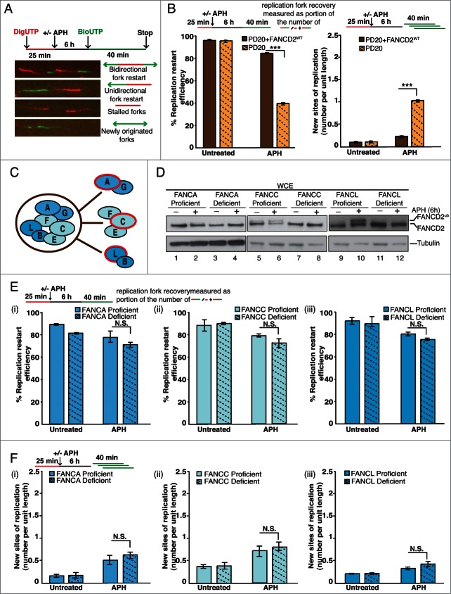
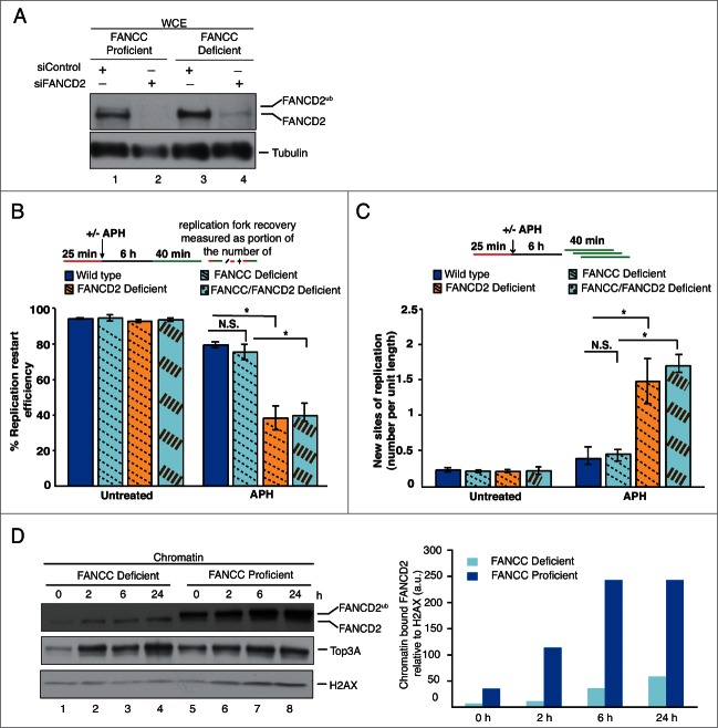
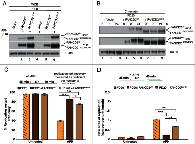
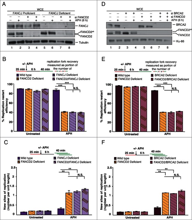
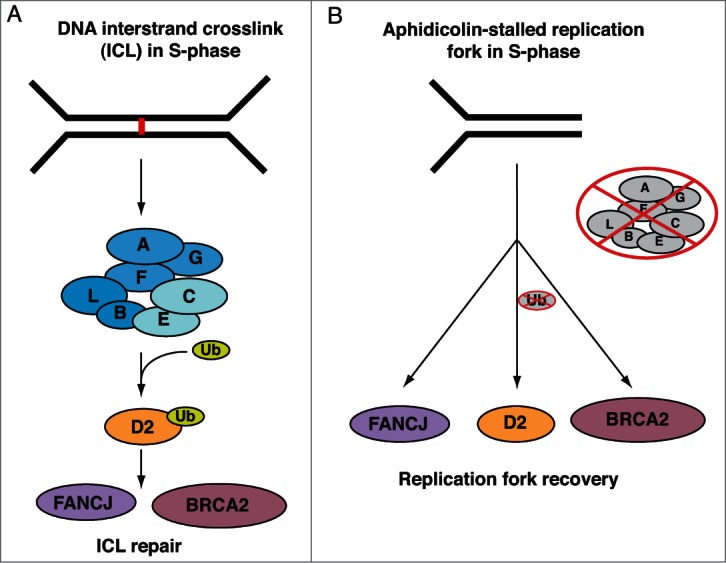
Fanconi Anemia (FA) is an inherited multi-gene cancer predisposition syndrome that is characterized on the cellular level by a hypersensitivity to DNA interstrand crosslinks (ICLs). To repair these lesions, the FA pathway proteins are thought to act in a linear hierarchy: Following ICL detection, an upstream FA core complex monoubiquitinates the central FA pathway members FANCD2 and FANCI, followed by their recruitment to chromatin. Chromatin-bound monoubiquitinated FANCD2 and FANCI subsequently coordinate DNA repair factors including the downstream FA pathway members FANCJ and FANCD1/BRCA2 to repair the DNA ICL. Importantly, we recently showed that FANCD2 has additional independent roles: it binds chromatin and acts in concert with the BLM helicase complex to promote the restart of aphidicolin (APH)-stalled replication forks, while suppressing the firing of new replication origins. Here, we show that FANCD2 fulfills these roles independently of the FA core complex-mediated monoubiquitination step. Following APH treatment, nonubiquitinated FANCD2 accumulates on chromatin, recruits the BLM complex, and promotes robust replication fork recovery regardless of the absence or presence of a functional FA core complex. In contrast, the downstream FA pathway members FANCJ and BRCA2 share FANCD2's role in replication fork restart and the suppression of new origin firing. Our results support a non-linear FA pathway model at stalled replication forks, where the nonubiquitinated FANCD2 isoform - in concert with FANCJ and BRCA2 - fulfills a specific function in promoting efficient replication fork recovery independently of the FA core complex.

Keywords: FA pathway; FANCD1/BRCA2; FANCD2; FANCD2 monoubiquitination; FANCJ; Fanconi Anemia; replication fork recovery.


Figures

Figure 1
Figure 1
(See previous page). The 3 FA core subcomplexes are dispensable for the restart of APH-stalled replication forks. (A) Representative images of DNA fibers accompanied with a schematic of defining sites of replication. Red tracts: DigU; green tracts: BioU. (B) Left panel: Replication fork restart efficiencies were compared between FANCD2-proficient (PD20+FANCD2WT) and -deficient (PD20) cells. Replication restart efficiency was measured as the number of restarted replication forks after APH-mediated fork stalling (DigU-BioU tracts), compared with the total number of DigU-labeled tracts (DigU + DigU-BioU). Right panel: The number of new sites of replication originating during the 40 min recovery period after APH treatment was compared between PD20+FANCD2WT and PD20 cells. New origins of replication were measured as the number of green-only (BioU) tracts per unit length. [N.B. The same DNA fiber assay analysis described in Fig. 1B for analyzing replication fork restart and new origin firing following APH treatment was used throughout this study.] (C) Schematic representation of the 3 FA core subcomplexes: FANCA-FANCG, FANCC-FANCE-FANCF, and FANCB-FANCL. FA patient-derived cell lines lacking FANCA, FANCC, or FANCL (proteins circled in red) were used to represent cells deficient for the respective FA core subcomplex. (D) WCEs from untreated or APH-treated, isogenic cell pairs that were either proficient or deficient for FANCA (lanes 1-4), FANCC (lanes 5-8) or FANCL (lanes 9-12) were analyzed for the presence of FANCD2 and FANCD2Ub. Tubulin, loading control. (E) Replication fork restart efficiencies after APH treatment was compared between isogenic cell pairs proficient or deficient for (i) FANCA, (ii) FANCC or (iii) FANCL. (F) The number of new replication sites originating during BioU labeling after APH treatment was compared between isogenic cell pairs proficient or deficient for (i) FANCA, (ii) FANCC or (iii) FANCL.
Figure 2.
Figure 2.
The nonubiquitinated FANCD2 isoform promotes the recovery of APH-stalled forks in FA core complex-deficient cells. (A) WCEs showing the efficiency of siRNA-mediated FANCD2 knockdown in FANCC-proficient or -deficient cells. Generated cell types: Wild type (PD331+C, siControl), FANCC-deficient (PD331, siControl), FANCD2-deficient (PD331+C, siFANCD2) and FANCC/FANCD2 double-deficient (PD331, siFANCD2). Tubulin, loading control. (B) Replication fork restart efficiencies after APH treatment were compared between the 4 cell types described in (A). (C) The number of new replication sites originating during BioU labeling after APH treatment was compared between the 4 cell types described in (A). (D) Left panel: FANCC-deficient or -proficient cells were either untreated or treated with APH for the indicated time points. Chromatin fractions isolated from these cells were analyzed for the presence of FANCD2, FANCD2Ub and Top3A. H2AX, loading control. Right panel: Immunoblot signals for FANCD2 shown the in left panel were analyzed by densitometry and normalized against H2AX signals using ImageJ.
Figure 3.
Figure 3.
FANCD2K561R binds chromatin and promotes restart of APH-stalled replication forks. (A) FANCD2-deficient cells (PD20) complemented with either empty vector, FANCD2WT or FANCD2K561R were either untreated or treated with APH, and analyzed for FANCD2 and FANCD2Ub. Ku-86, loading control. (B) The 3 cell types described in (A) were either untreated or treated with APH for 6h or 24h, and chromatin fractions from the cells were analyzed for the presence of FANCD2 and FANCD2Ub. Ku-86, loading control. (C) Replication fork restart efficiencies after APH treatment were compared between the 3 cell types described in (A). (D) The number of new replication sites originating during BioU labeling after APH treatment was compared between the 3 cell types described in (A).
Figure 4.
Figure 4.
FA pathway members FANCJ and BRCA2 cooperate with FANCD2 for the recovery of APH-stalled replication forks. (A) WCEs showing the efficiency of siRNA-mediated FANCD2 knockdown in FANCJ-proficient or -deficient cells. Generated cell types: Wild type (752/1T+J, siControl), FANCD2-deficient (752/1T+J, siFANCD2), FANCJ-deficient (752/1T, siControl) and FANCD2/FANCJ double- deficient (752/1T, siFANCD2). All 4 cell lines were untreated or treated with APH, and WCEs from these cells were analyzed for the presence of FANCJ, FANCD2 and FANCD2Ub. Tubulin, loading control. (B) Replication fork restart efficiencies after APH treatment were compared between wild type, FANCD2-, FANCJ- and FANCD2/FANCJ double-deficient cells (C) The number of new replication sites originating during BioU labeling after APH treatment was compared between Wildtype, FANCD2-, FANCJ- and FANCD2/FANCJ double-deficient cells. (D) WCEs showing the efficiency of siRNA-mediated FANCD2 and BRCA2 knockdown in wild type (PD331+C) cells. Generated cell types: wild type (PD331+C, siControl), FANCD2-deficient (PD331+C, siFANCD2), BRCA2-deficient (PD331+C, siBRCA2) and FANCD2/BRCA2 double-deficient (PD331+C, siFANCD2/siBRCA2). All 4 cell lines were untreated or treated with APH, and WCEs from these cells were analyzed for the presence of BRCA2, FANCD2 and FANCD2Ub. Ku-86: loading control. (E) Replication fork restart efficiencies after APH treatment were compared between wild type, FANCD2-, BRCA2- and FANCD2/BRCA2 double-deficient cells. (F) The number of new replication sites originating during BioU labeling after APH treatment was compared between wild type, FANCD2-, BRCA2- and FANCD2/BRCA2 double-deficient cells.
Figure 5.
Figure 5.
Distinct FA pathway models during DNA ICL repair vs. the restart of an APH-stalled replication fork. (A) Linear FA pathway model during DNA ICL repair. When replication forks converge at a DNA ICL (represented by a red line in the figure), the upstream FA core complex is activated and monoubiquitinates FANCD2, followed by its recruitment to the ICL on chromatin. Chromatin-bound FANCD2Ub then coordinates the actions of downstream DNA repair factors, including FANCJ and BRCA2, to facilitate ICL repair. (B) Non-linear FA pathway model during the recovery of APH-stalled replication forks. When a single moving replication fork is temporarily stalled by APH-treatment, nonubiquitinated FANCD2 is recruited to chromatin independently of the FA core complex. Chromatin-bound nonubiquitinated FANCD2 then functions in concert with FANCJ and BRCA2 to promote replication fork restart.

References

    1. Kee Y, D'Andrea AD. Molecular pathogenesis and clinical management of Fanconi anemia. J Clin Invest 2012; 122:3799-806; PMID:23114602; http://dx.doi.org/ 10.1172/JCI58321 - DOI - PMC - PubMed
    1. Kupfer GM. Fanconi anemia: a signal transduction and DNA repair pathway. Yale J Biol Med 2013; 86:491-7; PMID:24348213 - PMC - PubMed
    1. Naim V, Rosselli F. The FANC pathway and BLM collaborate during mitosis to prevent micro-nucleation and chromosome abnormalities. Nat Cell Biol 2009; 11:761-8; PMID:19465921; http://dx.doi.org/ 10.1038/ncb1883 - DOI - PubMed
    1. Kee Y, D'Andrea AD. Expanded roles of the Fanconi anemia pathway in preserving genomic stability. Genes Dev 2010; 24:1680-94; PMID:20713514; http://dx.doi.org/ 10.1101/gad.1955310 - DOI - PMC - PubMed
    1. Schlacher K, Wu H, Jasin M. A distinct replication fork protection pathway connects fanconi anemia tumor suppressors to RAD51-BRCA1/2. Cancer Cell 2012; 22:106-16; PMID:22789542; http://dx.doi.org/ 10.1016/j.ccr.2012.05.015 - DOI - PMC - PubMed

Publication types

MeSH terms