Damage-induced BRCA1 phosphorylation by Chk2 contributes to the timing of end resection
- PMID: 25659039
- PMCID: PMC4353239
- DOI: 10.4161/15384101.2014.972901
Damage-induced BRCA1 phosphorylation by Chk2 contributes to the timing of end resection
Abstract
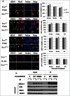
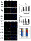
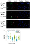
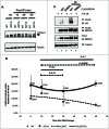
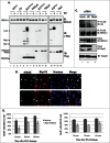
The BRCA1 tumor suppressor plays an important role in homologous recombination (HR)-mediated DNA double-strand-break (DSB) repair. BRCA1 is phosphorylated by Chk2 kinase upon γ-irradiation, but the role of Chk2 phosphorylation is not understood. Here, we report that abrogation of Chk2 phosphorylation on BRCA1 delays end resection and the dispersion of BRCA1 from DSBs but does not affect the assembly of Mre11/Rad50/NBS1 (MRN) and CtIP at DSBs. Moreover, we show that BRCA1 is ubiquitinated by SCF(Skp2) and that abrogation of Chk2 phosphorylation impairs its ubiquitination. Our study suggests that BRCA1 is more than a scaffold protein to assemble HR repair proteins at DSBs, but that Chk2 phosphorylation of BRCA1 also serves as a built-in clock for HR repair of DSBs. BRCA1 is known to inhibit Mre11 nuclease activity. SCF(Skp2) activity appears at late G1 and peaks at S/G2, and is known to ubiquitinate phosphodegron motifs. The removal of BRCA1 from DSBs by SCF(Skp2)-mediated degradation terminates BRCA1-mediated inhibition of Mre11 nuclease activity, allowing for end resection and restricting the initiation of HR to the S/G2 phases of the cell cycle.
Keywords: BRCA1; BRCA1, Breast Cancer Susceptibility Gene 1; DNA double-strand break repair; DSB, Double-Strand Break; HR, homologous Recombination; MRN, Mre11-Rad50-Nbs1 complex; NHEJ, Non-Homologous End Joining; SCF; SCF, Skp1-Cul1-F box protein complex; cell cycle; end-resection.
Figures


Similar articles
-
CSB interacts with BRCA1 in late S/G2 to promote MRN- and CtIP-mediated DNA end resection.Nucleic Acids Res. 2019 Nov 18;47(20):10678-10692. doi: 10.1093/nar/gkz784. Nucleic Acids Res. 2019. PMID: 31501894 Free PMC article.
-
DNA end resection is needed for the repair of complex lesions in G1-phase human cells.Cell Cycle. 2014;13(16):2509-16. doi: 10.4161/15384101.2015.941743. Cell Cycle. 2014. PMID: 25486192 Free PMC article.
-
Cell cycle-dependent complex formation of BRCA1.CtIP.MRN is important for DNA double-strand break repair.J Biol Chem. 2008 Mar 21;283(12):7713-20. doi: 10.1074/jbc.M710245200. Epub 2008 Jan 2. J Biol Chem. 2008. PMID: 18171670
-
Human syndromes with genomic instability and multiprotein machines that repair DNA double-strand breaks.Histol Histopathol. 2003 Jan;18(1):225-43. doi: 10.14670/HH-18.225. Histol Histopathol. 2003. PMID: 12507302 Review.
-
Good timing in the cell cycle for precise DNA repair by BRCA1.Cell Cycle. 2005 Sep;4(9):1216-22. doi: 10.4161/cc.4.9.2027. Epub 2005 Sep 13. Cell Cycle. 2005. PMID: 16103751 Review.
Cited by
-
Modulation of BRCA1 mediated DNA damage repair by deregulated ER-α signaling in breast cancers.Am J Cancer Res. 2022 Jan 15;12(1):17-47. eCollection 2022. Am J Cancer Res. 2022. PMID: 35141003 Free PMC article.
-
Phosphorylation of BRCA1 by ATM upon double-strand breaks impacts ATM function in end-resection: A potential feedback loop.iScience. 2022 Aug 15;25(9):104944. doi: 10.1016/j.isci.2022.104944. eCollection 2022 Sep 16. iScience. 2022. PMID: 36065181 Free PMC article.
-
DNA-damage-induced degradation of EXO1 exonuclease limits DNA end resection to ensure accurate DNA repair.J Biol Chem. 2017 Jun 30;292(26):10779-10790. doi: 10.1074/jbc.M116.772475. Epub 2017 May 17. J Biol Chem. 2017. PMID: 28515316 Free PMC article.
-
Hsp90: A New Player in DNA Repair?Biomolecules. 2015 Oct 16;5(4):2589-618. doi: 10.3390/biom5042589. Biomolecules. 2015. PMID: 26501335 Free PMC article. Review.
-
Wwox Binding to the Murine Brca1-BRCT Domain Regulates Timing of Brip1 and CtIP Phospho-Protein Interactions with This Domain at DNA Double-Strand Breaks, and Repair Pathway Choice.Int J Mol Sci. 2022 Mar 28;23(7):3729. doi: 10.3390/ijms23073729. Int J Mol Sci. 2022. PMID: 35409089 Free PMC article.
References
-
- Zhang J, Powell SN. The role of the BRCA1 tumor suppressor in DNA double-strand break repair. Mol Cancer Res 2005; 3:531-539; PMID:16254187; http://dx.doi.org/10.1158/1541-7786.MCR-05-0192 - DOI - PubMed
-
- Huen MSY, Shirley MHS, Chen J. BRCA1 and its toolbox for the maintenance of genome integrity. Nat Rev Mole Cell Biol 2010; 11:139-148; http://dx.doi.org/10.1038/nrm2831 - DOI - PMC - PubMed
-
- Scully R, Chen J, Ochs RL, Keegan K, Hoekstra M, Feunteun J, Livingston DM. Dynamic changes of BRCA1 subnuclear location and phosphorylation state are initiated by DNA damage. Cell 1997; 90:425-435; PMID:9267023; http://dx.doi.org/10.1016/S0092-8674(00)80503-6. - DOI - PubMed
-
- Cortez D, Wang Y, Qin J, Elledge SJ. Requirement of ATM-dependent phosphorylation of Brca1 in the DNA damage response to double-strand breaks. Science 1999; 286:1162-1166; PMID:10550055; http://dx.doi.org/10.1126/science.286.5442.1162. - DOI - PubMed
-
- Lee J-S, Collins KM, Brown AL, Lee C-H, Chung JH. hCds1-mediated phosphorylation of BRCA1 regulates the DNA damage response. Nature 2000; 404:201-204; PMID:10724175; http://dx.doi.org/10.1038/35004614. - DOI - PubMed
Publication types
MeSH terms
Substances
Grants and funding
LinkOut - more resources
Full Text Sources
Other Literature Sources
Molecular Biology Databases
Research Materials
Miscellaneous
