. 2015 Feb 6;10(2):e0118072.
doi: 10.1371/journal.pone.0118072. eCollection 2015.

Evaluation of resting state networks in patients with gliomas: connectivity changes in the unaffected side and its relation to cognitive function

Affiliations

Evaluation of resting state networks in patients with gliomas: connectivity changes in the unaffected side and its relation to cognitive function

Satoshi Maesawa et al. PLoS One. .

Abstract

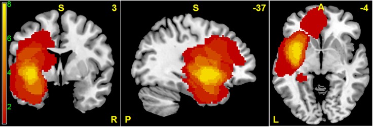
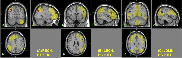
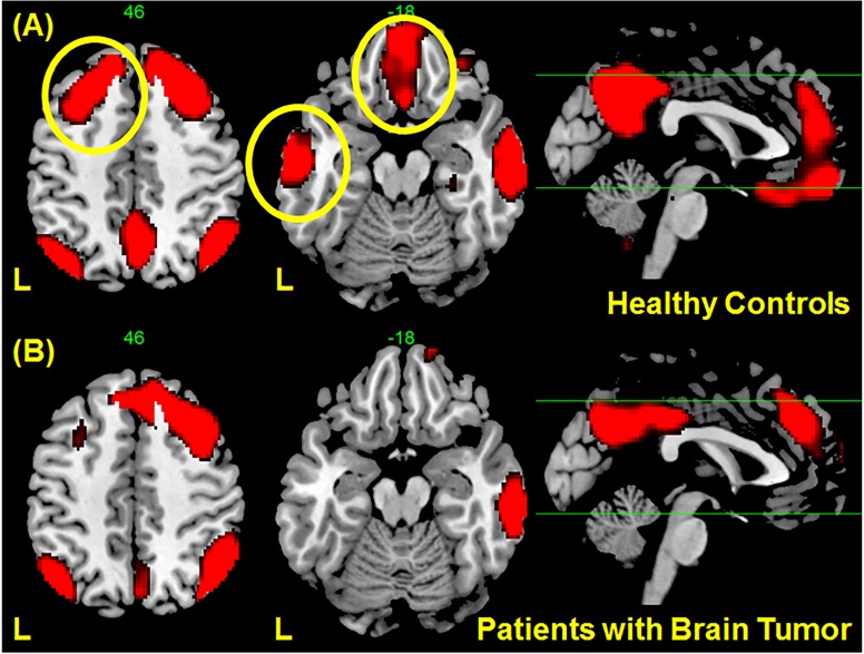
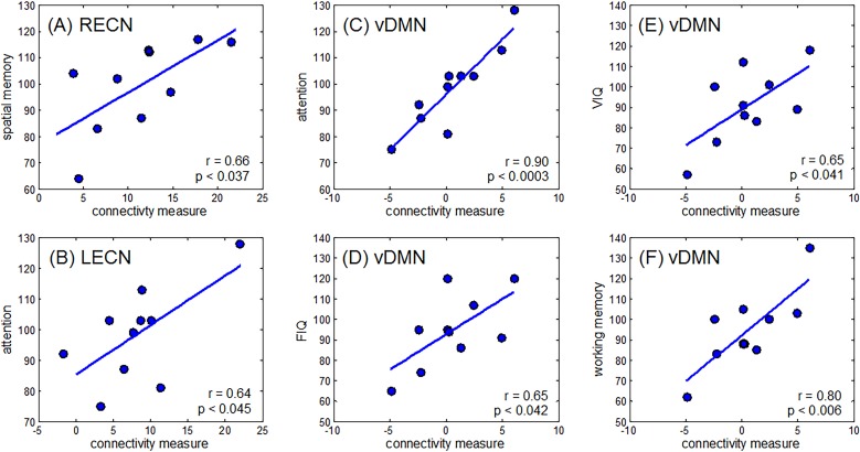
In this study, we investigated changes in resting state networks (RSNs) in patients with gliomas located in the left hemisphere and its relation to cognitive function. We hypothesized that long distance connection, especially between hemispheres, would be affected by the presence of the tumor. We further hypothesized that these changes would correlate with, or reflect cognitive changes observed in patients with gliomas. Resting state functional MRI datasets from 12 patients and 12 healthy controls were used in the analysis. The tumor's effect on three well-known RSNs including the default mode network (DMN), executive control network (ECN), and salience network (SN) identified using independent component analysis were investigated using dual regression analysis. Scores of neuropsychometric testing (WAIS-III and WMS-R) were also compared. Compared to the healthy control group, the patient group showed significant decrease in functional connectivity in the right angular gyrus/inferior parietal lobe of the ventral DMN and in the dorsolateral prefrontal cortex of the left ECN, whereas a significant increase in connectivity in the right ECN was observed in the right parietal lobe. Changes in connectivity in the right ECN correlated with spatial memory, while that on the left ECN correlated with attention. Connectivity changes in the ventral DMN correlated with attention, working memory, full IQ, and verbal IQ measures. Although the tumors were localized in the left side of the brain, changes in connectivity were observed in the contralateral side. Moreover, these changes correlated with some aspects of cognitive function indicating that patients with gliomas may undergo cognitive changes even in the absence of or before the onset of major symptoms. Evaluation of resting state networks could be helpful in advancing our hodological understanding of brain function in glioma cases.


Conflict of interest statement

Competing Interests: The authors have declared that no competing interests exist.

Figures

Fig 1
Fig 1. Spatial distribution of tumors in MNI standard space superimposed on a standard anatomical template.
This frequency map shows the number of patients with tumor in a given voxel.
Fig 2
Fig 2. Changes in functional connectivity in resting state networks.
Regions showing significant changes in functional connectivity in the (A) right executive control network (RECN), (B) left executive control network (LECN), and (C) ventral default mode network (vDMN) (p < 0.05 TFCE-cluster corrected for multiple comparison). HC–healthy control, BT–patients with brain tumor. Resting state networks are shown in yellow. Center of the cursor locates maximum significance.
Fig 3
Fig 3. Regions correlated with the right angular gyrus (Fig. 2C) obtained using seed-based connectivity analysis in (A) healthy controls and (B) patients with gliomas.
Correlations in several regions in the left hemisphere, indicated by yellow circle in (A), are no longer significant in patients with gliomas although only the region in the middle temporal gyrus showed statistical significance. The maps showed voxels with t values < 3.5.
Fig 4
Fig 4. Relationship between changes in connectivity and several cognitive measures.
Connectivity measures were extracted from regions shown in Fig. 2. Strong correlations were observed between changes in (A) RECN and spatial memory, (B) LECN and attention, (C) vDMN and attention, (D) vDMN and FIQ, (E) vDMN and VIQ, and (F) vDMN and working memory.

References

    1. Mahaley MS, Mettlin C, Natarajan N, Laws ER, Peace BB (1989) National survey of patterns of care for brain-tumor patients. J Neurosurg 71: 826–836. 10.3171/jns.1989.71.6.0826 - DOI - PubMed
    1. Sporns O (2011) The human connectome: A complex network. Ann N Y Acad Sci 1224: 109–125. 10.1111/j.1749-6632.2010.05888.x - DOI - PubMed
    1. Greicius MD, Krasnow B, Reiss AL, Menon V (2003) Functional connectivity in the resting brain: a network analysis of the default mode hypothesis. Proc Natl Acad Sci U S A 100: 253–258. 10.1073/pnas.0135058100 - DOI - PMC - PubMed
    1. Damoiseaux JS, Rombouts SARB, Barkhof F, Scheltens P, Stam CJ, et al. (2006) Consistent resting-state networks across healthy subjects. Proc Natl Acad Sci U S A 103: 13848–13853. 10.1073/pnas.0601417103 - DOI - PMC - PubMed
    1. Chen S, Ross TJ, Zhan W, Myers CS, Chuang KS, et al. (2008) Group independent component analysis reveals consistent resting-state networks across multiple sessions. Brain Res 1239: 141–151. 10.1016/j.brainres.2008.08.028 - DOI - PMC - PubMed

Publication types

LinkOut - more resources