Review
. 2015 Feb 6;5(3):a023119.
doi: 10.1101/cshperspect.a023119.

Genetic considerations in recurrent pregnancy loss

Affiliations
Review

Genetic considerations in recurrent pregnancy loss

Kassie J Hyde et al. Cold Spring Harb Perspect Med. .

Abstract

Human reproduction is remarkably inefficient; nearly 70% of human conceptions do not survive to live birth. Spontaneous fetal aneuploidy is the most common cause for spontaneous loss, particularly in the first trimester of pregnancy. Although losses owing to de novo fetal aneuploidy occur at similar frequencies among women with sporadic and recurrent losses, some couples with recurrent pregnancy loss have additional associated genetic factors and some have nongenetic etiologies. Genetic testing of the products of conception from couples experiencing two or more losses may aid in defining the underlying etiology and in counseling patients about prognosis in a subsequent pregnancy. Parental karyotyping of couples who have experienced recurrent pregnancy loss (RPL) will detect some couples with an increased likelihood of recurrent fetal aneuploidy; this may direct interventions. The utility of preimplantation genetic analysis in couples with RPL is unproven, but new approaches to this testing show great promise.


Figures

Figure 1.
Figure 1.
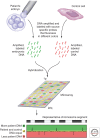
Analysis of blastomere DNA using array comparative genomic hybridization (aCGH). DNA microarrays allow comparison of the amounts of specific segments of DNA within a test sample to that from a control sample. The portion of the genome tested by a given microarray depends on the specific pieces of DNA contained in the spots on the array. For complete genomic hybridization, these spots represent the entire genome, from the beginning of the short arm of chromosome 1 (1p) to the end of the long arm of chromosome 22 plus the X and Y chromosomes. In aCGH, DNAs from a single test cell (e.g., a blastomere) and from a control cell are amplified and labeled with colored fluorescent probes (each DNA source with its own color). Equal amounts of DNA from each source are hybridized to the array chip displaying the specific segments of DNA being tested. A computer then reads the chip and plots out the colors seen in each dot of the array. When one sample is labeled red and the other green, spots at which the DNA from each source is present in equal amounts will fluoresce yellow. Imbalances in the amounts of DNA will be seen as red or green. For example, in this diagram, red fluorescence indicates a less patient DNA than in the control. Areas of balance and imbalance can be mapped across the complete genome.
Figure 2.
Figure 2.
Cytogenetic analysis of blastomere DNA using fluorescence in situ hybridization (FISH). DNA from a single cell (e.g., a blastomere) is collected at metaphase and the chromosomes are affixed to a surface for labeling and microscopic examination. Fluorescently labeled probes specific to known segments of a given chromosome are used to label the prepared chromosomes. Multicolor FISH uses between three and 11 differently colored probes to distinguish specific chromosomes. Gain or loss of the segment of a given chromosome labeled by a probe can be determined by simply counting the number of times a given color labels the DNA within the metaphase spread.
Figure 3.
Figure 3.
Biopsy methods for preimplantation genetic analysis. There are several stages at which material can be collected for preimplantation genetic analysis. Collection at the earliest stage involves removal of a polar body after the completion of meiosis I or meiosis II. Depicted is collection after fertilization (two pronuclei) and extrusion of the second polar body. Blastomere biopsy at the cleavage stage is typically performed when the embryo contains six to eight blastomeres; one to two blastomeres are retrieved from a single embryo. Trophectoderm biopsy is most frequently performed just before spontaneous embryo hatching from the zona pellucida on day 5 postfertilization (pf) and retrieves several trophectoderm cells. All biopsy methods involve manipulation of the zona pellucida. Using mechanical, chemical or laser-assisted techniques, a breach in the zona pellucida is made at the time of collection in polar body and cleavage stage blastomere biopsies. With trophectoderm biopsy, the zona pellucida is thinned on approximately day 3 pf, creating an area for preferential herniation of the trophectoderm on day 5 pf, just before spontaneous hatching. Trophectoderm cells are typically retrieved using laser-assisted techniques.

References

    1. Adler A, Lee HL, McCulloh DH, Ampeloquio E, Clarke-Williams M, Wertz BH, Grifo J 2014. Blastocyst culture selects for euploid embryos: Comparison of blastomere and trophectoderm biopsies. Fertil Steril 28: 485–491. - PubMed
    1. Alberman E 1988. The epidemiology of repeated abortion. In Early pregnancy loss: Mechanisms and treatment, (ed. Beard RW and Sharp F), pp. 9–17 Springer-Verlag, New York.
    1. Aldrich CL, Stephenson MD, Karrison T, Odem RR, Branch DW, Scott JR, Schreiber JR, Ober C 2001. HLA-G genotypes and pregnancy outcome in couples with unexplained recurrent miscarriage. Mol Hum Reprod 7: 1167–1172. - PubMed
    1. Angell RR 1994. Aneuploidy in older women. Higher rates of aneuploidy in oocytes from older women. Hum Reprod 9: 1199–1200. - PubMed
    1. Asadpor U, Totonchi M, Sabbaghian M, Hoseinifar H, Akhound MR, Zari Moradi Sh, Haratian K, Sadighi Gilani MA, Gourabi H, Mohseni Meybodi A 2013. Ubiquitin-specific protease (USP26) gene alterations associated with male infertility and recurrent pregnancy loss (RPL) in Iranian infertile patients. J Assist Reprod Genet 30: 923–931. - PMC - PubMed

LinkOut - more resources