Review
. 2015 Feb 6;6(1):a017848.
doi: 10.1101/cshperspect.a017848.

Clinical Aspects of Adult Tuberculosis

Affiliations
Review

Clinical Aspects of Adult Tuberculosis

Robert Loddenkemper et al. Cold Spring Harb Perspect Med. .

Abstract

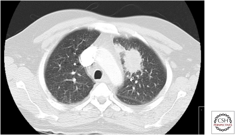
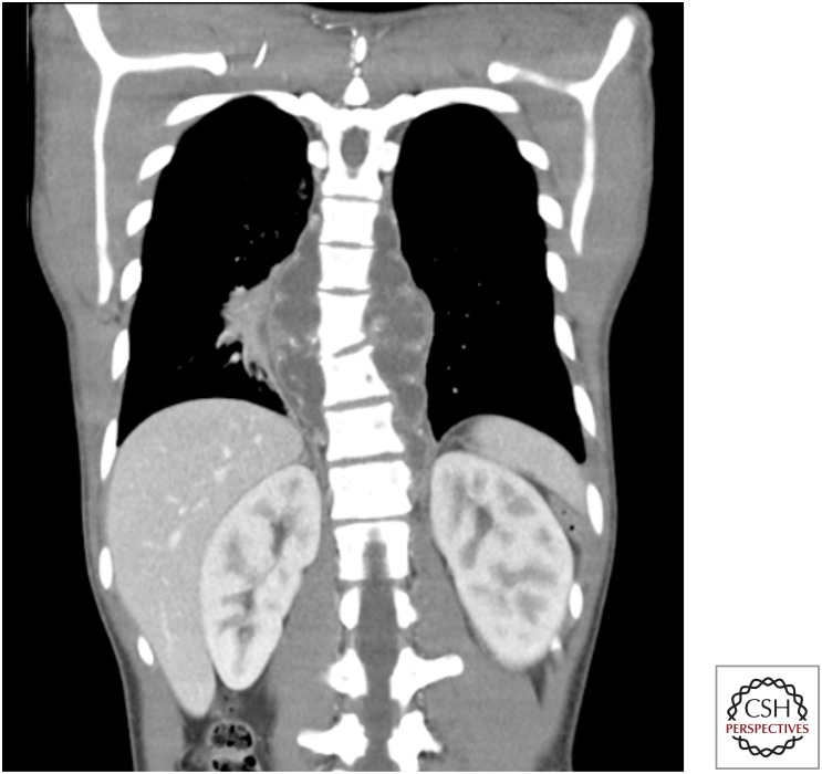
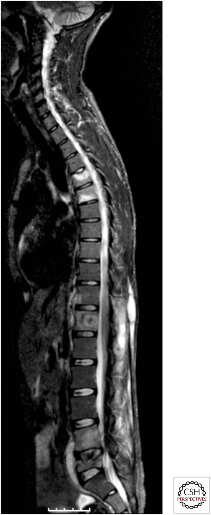
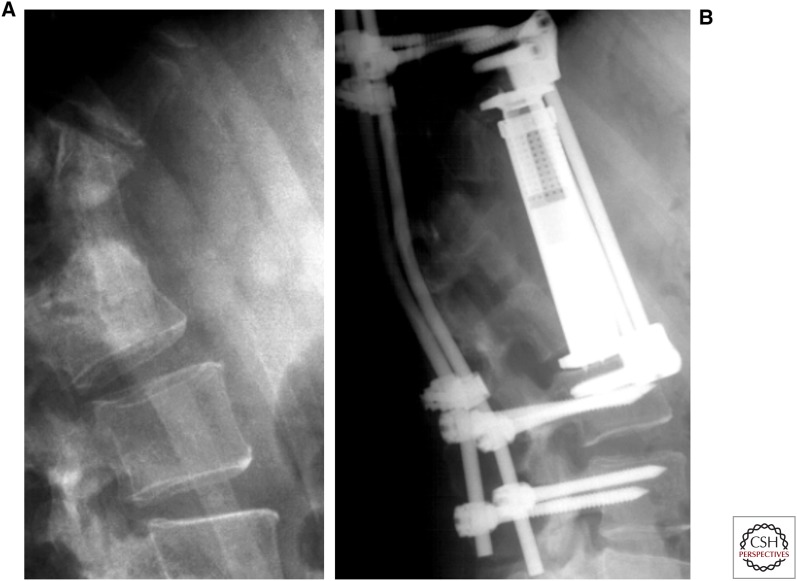
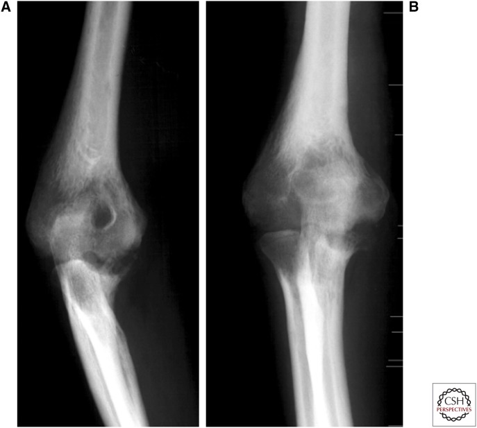
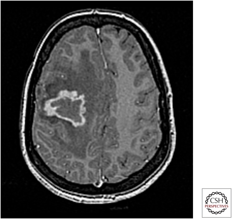
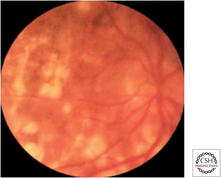
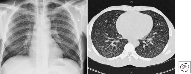
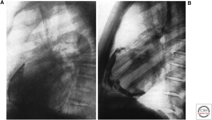
Tuberculosis (TB) in adults can present in a large number of ways. The lung is the predominant site of TB. Primary pulmonary TB should be distinguished from postprimary pulmonary TB, which is the most frequent TB manifestation in adults (70%-80% cases). Cough is common, although the chest radiograph often raises suspicion of disease. Sputum sampling is a key step in the diagnosis of TB, and invasive procedures such as bronchoscopy may be necessary to achieve adequate samples for diagnosis. Extrapulmonary involvement, which may present many years after exposure, occurs in a variable proportion of cases (20%-45%). This reflects the country of origin of patients and also the frequency of associated human immunodeficiency virus (HIV) coinfection. In the latter case, the presentation of TB is often nonspecific, and care needs to be taken to not miss the diagnosis. Anti-TB therapy should be given in line with proven (or assumed) drug resistance. In extrapulmonary TB, adjunctive therapeutic measures may be indicated; although in all cases, support is often required to ensure that people are able to complete treatment with minimal adverse events and maximal adherence to the prescribed regimen, and so reduce risk of future disease for themselves and others.


Figures

Figure 1.
Figure 1.
Ghon complex. (Figure reprinted from Fuehner et al. 2007, with permission, from Springer, © 2007.)
Figure 2.
Figure 2.
Disseminated BCG disease. Computed tomography (CT) of the chest showing bilateral patchy ground glass shadowing and consolidation in a patient who developed fevers, cough, and positive blood cultures for BCG following treatment with intravesical BCG for bladder carcinoma. (Figure provided by Marc Lipman.)
Figure 3.
Figure 3.
Cavitating tuberculosis presenting with hemoptysis (note arrows indicating cavities). (Figure reprinted from Fuehner et al. 2007, with permission, from Springer, © 2007.)
Figure 4.
Figure 4.
TB in the mucosa of the left main bronchus. Histology and culture from biopsy were positive for Mtb. (Figure provided by Robert Loddenkemper.)
Figure 5.
Figure 5.
Lung tuberculoma. Initially thought to be a primary lung cancer, bronchoscopic biopsy showed acid-fast bacilli on smear. Culture grew Mtb. (Figure provided by Marc Lipman.)
Figure 6.
Figure 6.
Age-specific female-to-male odds ratio (OR) of having extrapulmonary TB compared with pulmonary TB by site of extrapulmonary TB, Germany 1996–2000. Solid circle, lymphatic, peripheral; open circle, lymphatic intrathoracic; solid square, pleural; open square, genitourinary; solid triangle, osteoarticular; inverted solid triangle, meningeal; inverted open triangle, peritoneal. (Figure reprinted from Forssbohm et al. 2008, with permission, from the European Respiratory Society, © 2008.)
Figure 7.
Figure 7.
Silicotuberculosis. (Figure reprinted from Fuehner et al. 2007, with permission, from Springer, © 2007.)
Figure 8.
Figure 8.
Pneumonia of left upper lobe (lingula) caused by lymph node perforation. (Figure provided by Robert Loddenkemper.)
Figure 9.
Figure 9.
CT of the chest and bronchoscopy showing lymph node perforation, before and after treatment for TB. (Figure provided by Robert Loddenkemper.)
Figure 10.
Figure 10.
Extrathoracic lymph node disease. (Left figure, provided by the authors; right figure, reprinted from Siemon 2007, with permission, from Springer, © 2007.)
Figure 11.
Figure 11.
Unilateral large left pleural effusion attributable to tuberculosis. (Figure reprinted from Fuehner et al. 2007 with permission, from Springer, © 2007.)
Figure 12.
Figure 12.
Thoracoscopic image of early TB pleural effusion. Following drainage of 800 mL of serous effusion, typical miliary sago-like nodules in all parts of the right parietal pleura can be seen (1) on the diaphragm and (2) the anterior chest wall. (Figure reprinted from Loddenkemper et al. 2011, with permission, from Thieme, © 2011.)
Figure 13.
Figure 13.
Thoracoscopic image of established TB pleural effusion. Following drainage of 2000 mL of serous effusion, inflamed and hyperemic visceral and parietal pleura covered with fibrinous patches is evident. The posterior costovertebral region is shown with the diaphragm (1) and the chest wall (2). The lung cannot be visualized as it is fibrin-covered. The fibrinous adhesions pull the diaphragm cranially, producing tenting, and show vascularization at their base (arrows). (Figure reprinted from Loddenkemper et al. 2011, with permission, from Thieme, © 2011.)
Figure 14.
Figure 14.
(A) Combined histological and bacteriological yield (%) of standard techniques in diagnosis of TB-related pleural effusion. (B) Cultural yield (%) of standard techniques in diagnosis of TB-related pleural effusion. (Figure reprinted from Loddenkemper et al. 2011, with permission, from Thieme, © 2011.)
Figure 15.
Figure 15.
Tuberculous pleural empyema. (Figure reprinted from Siemon 2007, with permission, from Springer, © 2007.)
Figure 16.
Figure 16.
Coronal CT image of young male with back pain, fever, and weight loss with tuberculous paraspinal abscess secondary to vertebral involvement. (Figure provided by Marc Lipman.)
Figure 17.
Figure 17.
Spinal tuberculosis. Whole-spine MRI showing abnormal signal at multiple vertebral levels within cervical, thoracic, and lumbar spine, plus anterior paraspinal collections at sites of disease. (Figure provided by Marc Lipman.)
Figure 18.
Figure 18.
Spinal tuberculosis. (A) Destruction of vertebrae after tuberculous spondylitis; (B) treatment with external fixator. (Figure reprinted from Siemon 2007, with permission, from Springer, © 2007.)
Figure 19.
Figure 19.
Tuberculosis of the elbow joint. Bone loss is present at the distal humerus, its epidondyles, and also the olecranon and coronoidal process of the ulna. (Figure reprinted from Siemon 2007, with permission, from Springer, © 2007.)
Figure 20.
Figure 20.
Coronal MRI showing TB of the left ovary (black arrow from caudal). Right ovary (white arrow from lateral) with cystic enlarged follicles. (Figure reprinted from Siemon 2007, with permission, from Springer, © 2007.)
Figure 21.
Figure 21.
Splenic tuberculomas. (Figure reprinted from Siemon 2007, with permission, from Springer, © 2007.)
Figure 22.
Figure 22.
Abdominal TB attributable to TB. Pain and weight loss in an Indian male initially considered to result from disseminated abdominal malignancy. CT abdomen: (A) Pretreatment peritoneal and mesenteric thickening leading to omental “cake” (note oral contrast given); (B) posttreatment normal peritoneum and mesentery (no oral contrast given). (Figure provided by Marc Lipman.)
Figure 23.
Figure 23.
CNS tuberculoma. MRI brain with contrast shows enhancing lesion with associated edema and mass effect in a Somali female presenting with headache and a seizure. Biopsy-cultured Mtb (Figure provided by Marc Lipman.)
Figure 24.
Figure 24.
Retinal tuberculous lesions. (Figure reprinted from Siemon 2007, with permission, from Springer, © 2007.)
Figure 25.
Figure 25.
Miliary TB on chest radiograph and CT. (Figure reprinted from Fuehner et al. 2007, with permission, from Springer, © 2007.)
Figure 26.
Figure 26.
Tracheo-cutaneous fistula with scrofuloderma. (Figure reprinted from Siemon 2007, with permission, from Springer, © 2007.)
Figure 27.
Figure 27.
Tuberculous pericarditis with bilateral pleural effusions. (A) CT; (B) echocardiography. (Figure reprinted from Siemon 2007, with permission, from Springer, © 2007.)
Figure 28.
Figure 28.
Calcification after tuberculous pericarditis. (Figure reprinted from Uehlinger et al. 1959, with permission, from EMH Schweizer Ärzteverlag, © 1959.)

References

    1. Aït-Khaled N, Alarcón E, Armengol R, Bissell K, Boillot F, Caminero JA, Chiang C-Y, Clevenbergh P, Dlodlo R, Enarson DA, et al. 2010. Management of tuberculosis: A guide to the essentials of good practice. Paris, France, International Union Against Tuberculosis and Lung Disease (The Union) http://www.theunion.org/what-we-do/publications/technical/english/pub_or...
    1. Almadi MA, Ghosh S, Aljebreen AM. 2009. Differentiating intestinal tuberculosis from Crohn’s disease: A diagnostic challenge. Am J Gastroenterol 104: 1003–1012. - PubMed
    1. Anderson C, White J, Abubakar I, Lipman M, Tamne S, Anderson SR, deKoningh J, Dart S. 2014. Raising standards in UK TB control: Introducing cohort review. Thorax 69: 187–189. - PubMed
    1. Bernaerts A, Vanhoenacker FM, Parizel PM, Van Goethem JW, Van Altena R, Laridon A, De Roeck J, Coeman V, De Schepper AM. 2003. Tuberculosis of the central nervous system: Overview of neuroradiological findings. Eur Radiol 13: 1876–1890. - PubMed
    1. Bhat VK, Latha P, Upadhya D, Hegde J. 2009. Clinicopathological review of tubercular laryngitis in 32 cases of pulmonary Kochs. Am J Otolaryngol 30: 327–330. - PubMed

MeSH terms

Substances

LinkOut - more resources