. 2015 Apr 10;290(15):9500-10.
doi: 10.1074/jbc.M114.630871. Epub 2015 Feb 6.

The major autoantibody epitope on factor H in atypical hemolytic uremic syndrome is structurally different from its homologous site in factor H-related protein 1, supporting a novel model for induction of autoimmunity in this disease

Affiliations

The major autoantibody epitope on factor H in atypical hemolytic uremic syndrome is structurally different from its homologous site in factor H-related protein 1, supporting a novel model for induction of autoimmunity in this disease

Arnab Bhattacharjee et al. J Biol Chem. .

Abstract

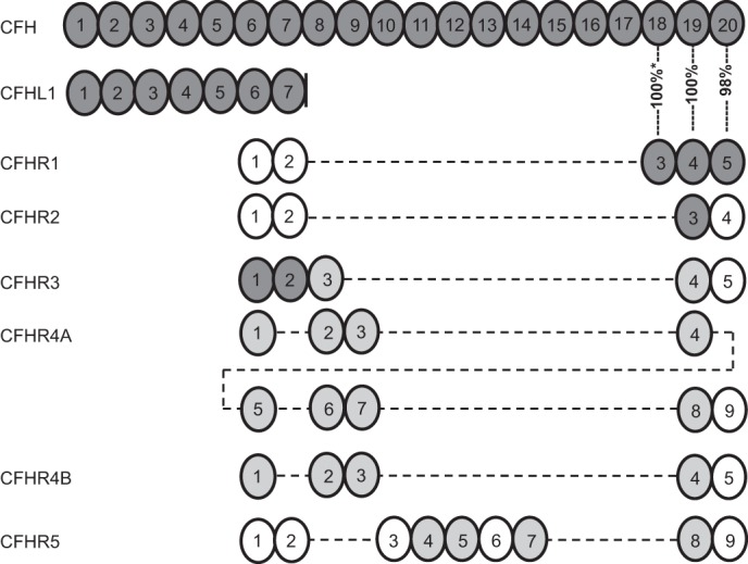
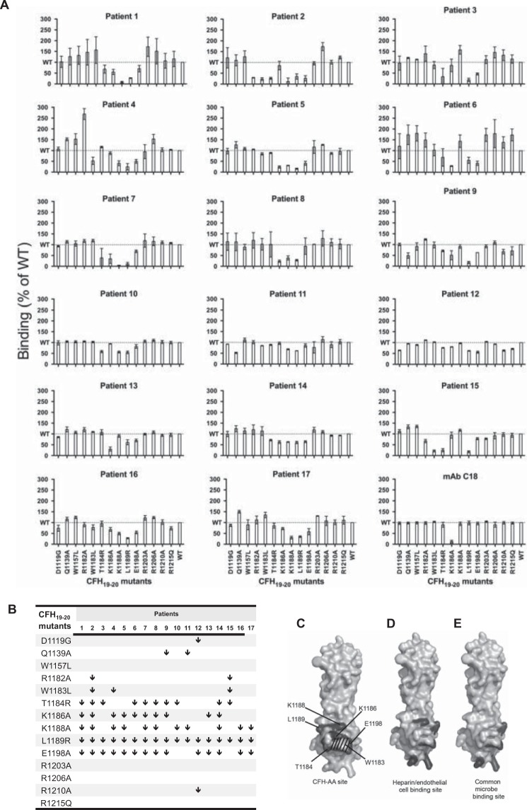
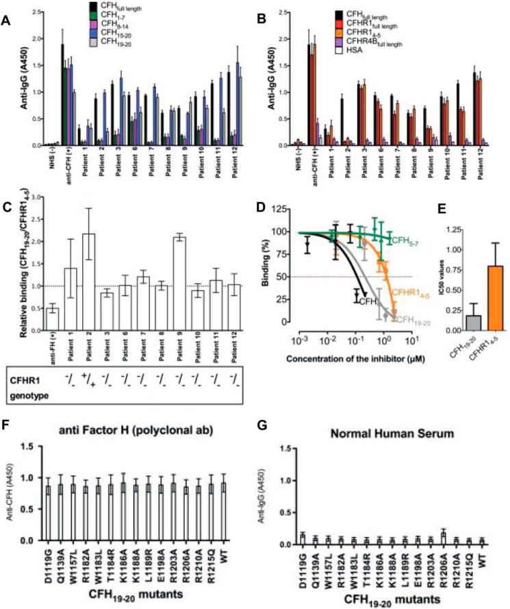
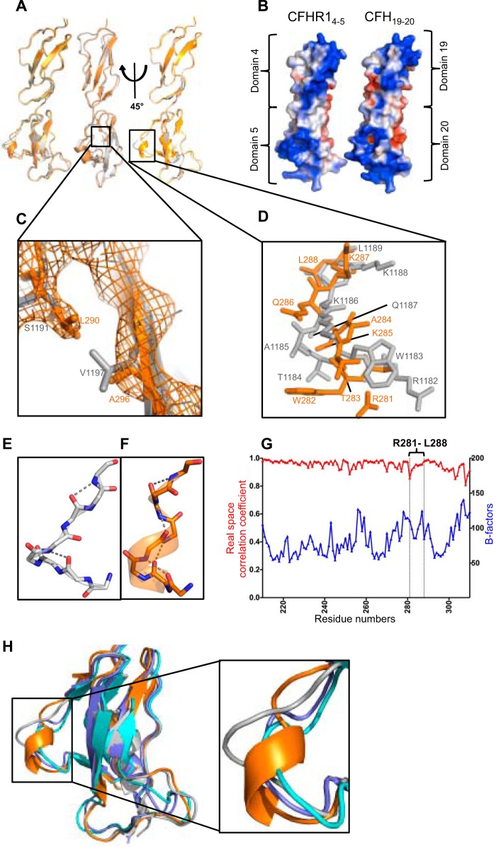
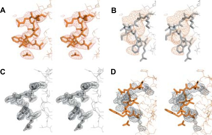
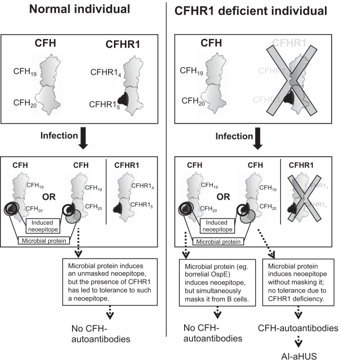
Atypical hemolytic uremic syndrome (aHUS) is characterized by complement attack against host cells due to mutations in complement proteins or autoantibodies against complement factor H (CFH). It is unknown why nearly all patients with autoimmune aHUS lack CFHR1 (CFH-related protein-1). These patients have autoantibodies against CFH domains 19 and 20 (CFH19-20), which are nearly identical to CFHR1 domains 4 and 5 (CFHR14-5). Here, binding site mapping of autoantibodies from 17 patients using mutant CFH19-20 constructs revealed an autoantibody epitope cluster within a loop on domain 20, next to the two buried residues that are different in CFH19-20 and CFHR14-5. The crystal structure of CFHR14-5 revealed a difference in conformation of the autoantigenic loop in the C-terminal domains of CFH and CFHR1, explaining the variation in binding of autoantibodies from some aHUS patients to CFH19-20 and CFHR14-5. The autoantigenic loop on CFH seems to be generally flexible, as its conformation in previously published structures of CFH19-20 bound to the microbial protein OspE and a sialic acid glycan is somewhat altered. Cumulatively, our data suggest that association of CFHR1 deficiency with autoimmune aHUS could be due to the structural difference between CFHR1 and the autoantigenic CFH epitope, suggesting a novel explanation for CFHR1 deficiency in the pathogenesis of autoimmune aHUS.

Keywords: Atypical Hemolytic Uremic Syndrome; Autoimmune Disease; Autoimmunity; Complement Regulation; Immunoglobulin G (IgG); Immunology; Structure-Function Study; Thrombotic Microangiopathy; X-ray Crystallography.


Figures

FIGURE 1.
FIGURE 1.
Schematic illustration indicating the amino acid sequence identity of CFH to other members of the CFH family. Each CFHR or CFHL domain is shown below the domain of CFH to which it has the highest amino acid sequence identity. For sequence identities of 32–49%, the domains are shown in white; for 57–84% identity, in light gray; and for 85–100% identity, in dark gray. The identity between domains 3–5 of CFHR1 and domains 18–20 of CFH is indicated as a percentage. The asterisk indicates that the sequence identity of domain 3 in the basic isoform of CFHR1 to domain 18 in CFH is 100%, whereas that of the acidic isoform is 95% (12). CFHL-1 (CFH-like molecule-1) is an alternatively spliced transcript from the CFH gene with four unique residues following domain 7.
FIGURE 2.
FIGURE 2.
Mapping of the CFH-AA-binding region on CFH19–20. A, binding of IgG from 17 aHUS patients and mAb C18 to 14 CFH19–20 constructs with various single-point mutations in relation to binding to WT CFH19–20. Error bars indicate S.E., and the level of WT binding is indicated by dotted lines. B, comparison of the autoantibody-binding epitopes by identification of the mutations that impaired binding of patient IgG by at least 30% (indicated by ↓). C, locations of the residues involved in binding of autoantibodies to CFH19–20 as indicated in dark gray and annotated on a previously published structure of CFH19–20 (Protein Data Bank code 2G7I) (7). The location of Trp-1183 is indicated by stripes. D and E, for comparison, the locations of residues involved in binding of CFH19–20 to heparin (5, 40) (D) and in the common microbe-binding site on CFH19–20 (30) (E) are indicated in dark gray.
FIGURE 3.
FIGURE 3.
Binding of autoantibodies from autoimmune aHUS patients to CFH and CFHR1. A, binding of autoantibodies to CFH (full-length) and its fragments CFH1–7, CFH8–14, CFH15–20, and CFH19–20. B, binding of IgG autoantibodies to CFH, CFHR1 (full-length), CFHR14–5, and CFHR4B (full-length). Human serum albumin (HSA) and/or normal human serum (NHS) was used as a negative control, and goat anti-CFH polyclonal antibody was used as a positive control. C, bar diagram elucidating the relative binding ratio of the patient autoantibodies to CFH19–20 and CFHR14–5. Error bars indicate S.E. The CFHR1 deficiency of each patient is shown below. D, the binding of purified IgG from patient 11 to CFH19–20 was tested in the presence of increasing concentrations of CFH19–20 or CFHR14–5. CFH5–7 was used as a negative control. E, bar diagram of the concentration of CFH19–20 or CFHR14–5 needed for 50% inhibition (IC50) obtained from three independent experiments performed in triplicates. F, binding of anti-CFH polyclonal antibody to the CFH19–20 mutants. G, no binding was noticed between CFH19–20 mutants and IgG from normal human serum. Error bars indicate S.E.
FIGURE 4.
FIGURE 4.
Crystal structure of CFHR14–5 and comparison with the previously solved structure of CFH19–20. A, structural superposition of the two molecules of CFHR14–5 (orange and yellow) found in the asymmetric unit along with CFH19–20 (gray) shown in a cartoon representation. B, comparison of the surface charge potentials of CFHR14–5 and CFH19–20. Potentials on the solvent-accessible surfaces were calculated and displayed at the ±2 kT/e level on both structures after modeling all of the missing side chains of the previously published structure of CFH19–20 (Protein Data Bank code 2G7I) (7). C, close-up view of the two residues that are different in the amino acid sequences of these two protein constructs with the 2mFo − DFc electron density map of CFHR14–5 shown. D, close-up view of the region in which the tertiary structures of CFHR14–5 and CFH19–20 are dissimilar (Arg-1182–Leu-1189 of CFH19–20 and the corresponding Arg-281–Leu-288 of CFHR14–5) with backbone and side chain atoms shown as a stick model. This region corresponds to the CFH-AA-binding site shown in Fig. 2C. The hydrogen bonds found in the autoantigenic loop of CFH (E) and its homologous region in CFHR1 (F) that stabilize the structure. G, comparison of the real space correlation constants and B-factors of the residues of CFHR14–5. The B-factors are indicated on the right y axis, and the real space correlation coefficients are indicated on the left y axis. The CFH-AA site (Arg-281–Leu-288) is indicated. H, cartoon representation of the structural superposition of CFHR14–5 (orange; Protein Data Bank code 4MUC) with CFH19–20 (gray; code 2G7I (7)), CFH19–20 in complex with a sialic acid glycan and C3d (slate; code 4ONT (22)), and CFH19–20 in complex with OspE (turquoise; code 4J38 (29)).
FIGURE 5.
FIGURE 5.
Comparison of the models and 2mFo − DFc electron density maps of the CFH-AA-binding site of CFH19–20 and the corresponding site of CFHR14–5. The CFHR14–5 loop region (Arg-281–Leu-288; A) and the CFH19–20 loop region (Arg-1182–Leu-1189; B) are shown with the CFHR14–5 2mFo − DFc electron density map. The CFH19–20 loop region (Arg-1182–Leu-1189; C) and the CFHR14–5 loop region (Arg-281–Leu-288; D) are shown with the CFH19–20 2mFo − DFc electron density map. A stick model and electron density map of CFHR14–5 are displayed in orange, and those of CFH19–20 in gray.
FIGURE 6.
FIGURE 6.
Schematic illustration of a model to explain the occurrence of CFH-AAs in aHUS, with special attention to the strong association of autoimmune aHUS with homozygous deficiency of CFHR1. Upper panels, the phenotypes of normal and CFHR1-deficient individuals are shown schematically to indicate the structural difference observed between the CFH-AA site and the corresponding site on CFHR1 (dark gray protrusion). In the novel “induced neoepitope model,” binding of a microbial protein to CFH domain 20 (adjacent to the autoantigenic Arg-1182–Leu-1189 loop, thus not masking it from B-cells) induces a conformational change in the Arg-1182–Leu-1189 loop of domain 20, thereby making its conformation similar to that of CFHR1 domain 5.

Similar articles

Cited by

References

    1. Noris M., Remuzzi G. (2009) Atypical hemolytic-uremic syndrome. N. Engl. J. Med. 361, 1676–1687 - PubMed
    1. Sánchez-Corral P., Melgosa M. (2010) Advances in understanding the aetiology of atypical haemolytic uraemic Syndrome. Br. J. Haematol 150, 529–542 - PubMed
    1. Zipfel P. F., Edey M., Heinen S., Józsi M., Richter H., Misselwitz J., Hoppe B., Routledge D., Strain L., Hughes A. E., Goodship J. A., Licht C., Goodship T. H., Skerka C. (2007) Deletion of complement factor H-related genes CFHR1 and CFHR3 is associated with atypical hemolytic uremic syndrome. PLoS Genet 3, e41. - PMC - PubMed
    1. Venables J. P., Strain L., Routledge D., Bourn D., Powell H. M., Warwicker P., Diaz-Torres M. L., Sampson A., Mead P., Webb M., Pirson Y., Jackson M. S., Hughes A., Wood K. M., Goodship J. A., Goodship T. H. (2006) Atypical haemolytic uraemic syndrome associated with a hybrid complement gene. PLoS Med. 3, e431. - PMC - PubMed
    1. Lehtinen M. J., Rops A. L., Isenman D. E., van der Vlag J., Jokiranta T. S. (2009) Mutations of factor H impair regulation of surface-bound C3b by three mechanisms in atypical hemolytic uremic syndrome. J. Biol. Chem. 284, 15650–15658 - PMC - PubMed

Publication types

MeSH terms

Associated data