

Noninvasive pulmonary artery wave intensity analysis in pulmonary hypertension
- PMID: 25659483
- PMCID: PMC4469876
- DOI: 10.1152/ajpheart.00480.2014
Noninvasive pulmonary artery wave intensity analysis in pulmonary hypertension
Abstract
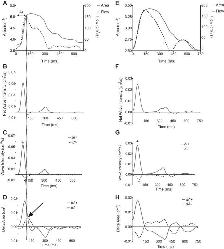
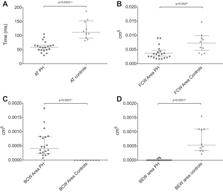
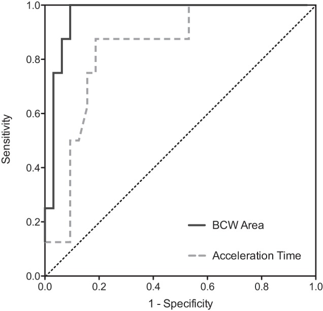
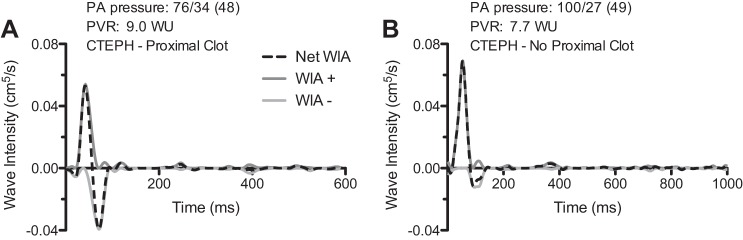
Pulmonary wave reflections are a potential hemodynamic biomarker for pulmonary hypertension (PH) and can be analyzed using wave intensity analysis (WIA). In this study we used pulmonary vessel area and flow obtained using cardiac magnetic resonance (CMR) to implement WIA noninvasively. We hypothesized that this method could detect differences in reflections in PH patients compared with healthy controls and could also differentiate certain PH subtypes. Twenty patients with PH (35% CTEPH and 75% female) and 10 healthy controls (60% female) were recruited. Right and left pulmonary artery (LPA and RPA) flow and area curves were acquired using self-gated golden-angle, spiral, phase-contrast CMR with a 10.5-ms temporal resolution. These data were used to perform WIA on patients and controls. The presence of a proximal clot in CTEPH patients was determined from contemporaneous computed tomography/angiographic data. A backwards-traveling compression wave (BCW) was present in both LPA and RPA of all PH patients but was absent in all controls (P = 6e(-8)). The area under the BCW was associated with a sensitivity of 100% [95% confidence interval (CI) 63-100%] and specificity of 91% (95% CI 75-98%) for the presence of a clot in the proximal PAs of patients with CTEPH. In conclusion, WIA metrics were significantly different between patients and controls; in particular, the presence of an early BCW was specifically associated with PH. The magnitude of the area under the BCW showed discriminatory capacity for the presence of proximal PA clot in patients with CTEPH. We believe that these results demonstrate that WIA could be used in the noninvasive assessment of PH.
Keywords: cardiac magnetic resonance imaging; hemodynamics; pulmonary hypertension; wave intensity.
Copyright © 2015 the American Physiological Society.
Figures
Comment in
-
Letter to the editor: Comparing pace and speed in the pulmonary circulation?Am J Physiol Heart Circ Physiol. 2016 Apr 1;310(7):H949. doi: 10.1152/ajpheart.00065.2016. Am J Physiol Heart Circ Physiol. 2016. PMID: 27036400 No abstract available.
-
Reply to: "Letter to the editor: Comparing pace and speed in the pulmonary circulation?".Am J Physiol Heart Circ Physiol. 2016 Apr 1;310(7):H950. doi: 10.1152/ajpheart.00120.2016. Am J Physiol Heart Circ Physiol. 2016. PMID: 27036401 No abstract available.
References
-
- Castelain V, Herve P, Lecarpentier Y, Duroux P, Simonneau G, Chemla D. Pulmonary artery pulse pressure and wave reflection in chronic pulmonary thromboembolism and primary pulmonary hypertension. J Am Coll Cardiol 37: 1085–1092, 2001. - PubMed
-
- Davies JE, Alastruey J, Francis DP, Hadjiloizou N, Whinnett ZI, Manisty CH, Aguado-Sierra J, Willson K, Foale RA, Malik IS, Hughes AD, Parker KH, Mayet J. Attenuation of wave reflection by wave entrapment creates a “horizon effect” in the human aorta. Hypertension 60: 778–785, 2012. - PubMed
-
- Davies JE, Whinnett ZI, Francis DP, Willson K, Foale RA, Malik IS, Hughes AD, Parker KH, Mayet J. Use of simultaneous pressure and velocity measurements to estimate arterial wave speed at a single site in humans. Am J Physiol Heart Circ Physiol 290: H878–H885, 2006. - PubMed
Publication types
MeSH terms
Grants and funding
LinkOut - more resources
Full Text Sources
Other Literature Sources
Medical
Miscellaneous